Titan Spine Appoints Chad Kolean as Chief Financial Officer

September 12, 2017

MEQUON, Wis.–(BUSINESS WIRE)–Titan Spine, a medical device surface technology company focused on developing innovative spinal interbody fusion implants, today announced the appointment of Chad Kolean as the Company’s Chief Financial Officer (CFO). In his role, Kolean will oversee Titan’s Finance Team and work to build forward-looking financial preparedness as the Company continues to experience rapid growth following the launch of its nanoLOCK® surface technology.

Prior to joining Titan, Mr. Kolean served as the CFO for Cellectar Biosciences, a life sciences company, where he drove a management restructuring and raised substantial funds resulting in uplisting to the NASDAQ Capital Market. Previously, he served as the CFO at Pioneer Surgical Technologies, providing the organization with leadership strategy, development and execution, Board support, investor relations, budgeting, forecasting, operations and regulatory due diligence. Kolean is a certified public accountant and earned his B.A. in Business Administration from Hope College.

Mr. Kolean commented, “I am thrilled to join the team at Titan Spine, particularly at such an exciting time for the company. We are well-positioned for continued financial growth that capitalizes on a strong first half of 2017. Titan Spine is making substantial progress addressing the key needs for both patients and surgeons, and the company’s growth is a direct reflection of the increased demand surgeons have for the advantages of our nanoLOCK® surface technology.”

Peter Ullrich, Chief Executive Officer of Titan Spine, added, “Chad’s appointment as our CFO validates and reflects our rapid expansion as we continue to disrupt our industry and redefine the role of interbody devices in the fusion process. The growing demand for our technology and our strong sales team has led to significant growth, especially in the first half of this year, and we believe Chad will help drive our continued progress, further benefiting patients at the nano-cellular level to heal faster following spinal fusions.”

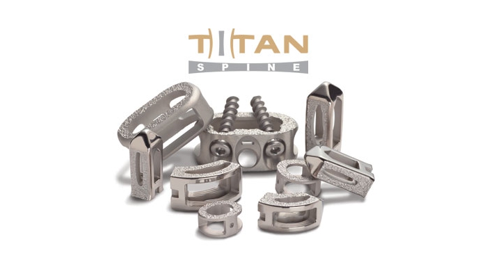
Titan Spine offers a full line of Endoskeleton® titanium implants that feature its proprietary nanoLOCK® surface technology, which was launched in the U.S. in October 2016 following FDA clearance in late 2014. The nanoLOCK® surface technology consists of a unique combination of roughened topographies at the macro, micro, and nano levels (MMN™). This unique combination of surface topographies is designed to create an optimal host-bone response and actively participate in the fusion process by promoting the upregulation of osteogenic and angiogenic factors necessary for bone growth, encouraging natural production of bone morphogenetic proteins (BMPs), downregulating inflammatory factors, and creating the potential for a faster and more robust fusion.2,3,4 All Endoskeleton® devices are covered by the company’s risk share warranty.

About Titan Spine

Titan Spine, LLC is a surface technology company focused on the design and manufacture of interbody fusion devices for the spine. The company is committed to advancing the science of surface engineering to enhance the treatment of various pathologies of the spine that require fusion. Titan Spine, located in Mequon, Wisconsin and Laichingen, Germany, markets a full line of Endoskeleton® interbody devices featuring its proprietary textured surface in the U.S. and portions of Europe through its sales force and a network of independent distributors. To learn more, visit www.titanspine.com.

1 Olivares-Navarrete, R., Hyzy S.L., Gittens, R.A., Berg, M.E., Schneider, J.M., Hotchkiss, K., Schwartz, Z., Boyan, B. D. Osteoblast lineage cells can discriminate microscale topographic features on titanium-aluminum-vanadium surfaces. Ann Biomed Eng. 2014 Dec; 42 (12): 2551-61.

Olivares-Navarrete, R., Hyzy, S.L., Slosar, P.J., Schneider, J.M., Schwartz, Z., and Boyan, B.D. (2015). Implant materials generate different peri-implant inflammatory factors: PEEK promotes fibrosis and micro-textured titanium promotes osteogenic factors. Spine, Volume 40, Issue 6, 399–404.

Olivares-Navarrete, R., Gittens, R.A., Schneider, J.M., Hyzy, S.L., Haithcock, D.A., Ullrich, P.F., Schwartz, Z., Boyan, B.D. (2012). Osteoblasts exhibit a more differentiated phenotype and increased bone morphogenetic production on titanium alloy substrates than poly-ether-ether-ketone. The Spine Journal, 12, 265-272.

4 Olivares-Navarrete, R., Hyzy, S.L., Gittens, R.A., Schneider, J.M., Haithcock, D.A., Ullrich, P.F., Slosar, P. J., Schwartz, Z., Boyan, B.D. (2013). Rough titanium alloys regulate

Contacts

Company:
Titan Spine
Andrew Shepherd, 866-822-7800
ashepherd@titanspine.com
or
Media:
The Ruth Group
Kirsten Thomas, 508-280-6592
kthomas@theruthgroup.com

Researchers at HSS Receive Grants to Study Bone Stress Injuries

NEW YORKSept. 12, 2017 /PRNewswire-USNewswire/ — Two research teams at Hospital for Special Surgery (HSS) have been awarded grants by the NBA and GE Healthcare Orthopedics and Sports Medicine Collaboration. This strategic partnership between NBA and GE Healthcare was launched to collaborate with leading clinical researchers to address the prevention, diagnosis and treatment of musculoskeletal injuries affecting NBA players and everyday athletes.

A team led by Brett Toresdahl, MD, primary care sports medicine physician at HSS, received a grant of nearly $300,000that will go towards investigating the use of ultrasound imaging to monitor healing and guide readiness of return to play in athletes with bone stress injuries.

Bone stress injuries of the lower leg and foot are common among elite basketball players and can require a long period of rest in order to fully recover. Determining when a player is ready to return to play has historically been difficult as it relies primarily on a player’s symptoms since signs of healing are not reliably seen with standard imaging (such as MRI and X-ray).

“There is little known about the connection between ultrasound findings, symptoms, and readiness for return to sport for athletes with bone stress injuries,” said Dr. Toresdahl, principle investigator of the study. “Our goal is to evaluate the use of serial ultrasound to assess bony healing to optimize treatment and more accurately predict readiness for return-to-play.”

The investigators will recruit adults between 18 and 50 years of age who have a bone stress injury in the lower leg or foot as a result of sport or exercise and diagnosed by MRI.

Participants will receive standard treatment of rest and crutches, splint and/or walking boot. An ultrasound will be performed every two weeks for 12 weeks at the site of the injury. Simultaneously, patients will be tracking their pain, activity and return to activity. A radiologist will grade the amount and appearance of bony callus formation and then a repeat MRI will be performed at 12 weeks.

“Our findings could provide doctors with a new way to guide athletes to safely return to play after a bone stress injury with a minimal risk of re-injury,” added Dr. Toresdahl.

The Athlete’s Foot

The other grant received by HSS went to a team led by Martin O’Malley, MD, foot and ankle surgeon at HSS.  This $100,000 grant will be used to assess risk factors and intervention strategies for fifth metatarsal stress fractures, the most common type of stress fractures for basketball players. These fractures can be difficult to treat, leading to long-term consequences for an elite basketball player.

“Currently, there is limited knowledge on how to identify athletes who are at a high risk for a fifth metatarsal stress fracture,” said Dr. O’Malley, principle investigator of this study. “Additionally, we don’t have a clinical understanding on how to intervene before such injury occurs.”

This research will utilize data from the screening of collegiate and professional basketball players to identify factors of foot structure that are common in those who sustained a fifth metatarsal injury.  The investigators will employ a specialized robotic device that will stimulate walking and jumping in cadavers to find features that increase the mechanical burden on this bone.

Testing will also be done to assess the ability of intervention strategies to reduce this mechanical burden and thus, the risk of injury.

“We hope to gain objective biomechanical evidence that will rationalize intervention strategies such as orthotic devices and targeted muscle activation in order to reduce risk of injury and re-injury,” added Dr. O’Malley.

About Hospital for Special Surgery
Hospital for Special Surgery (HSS) is the world’s leading academic medical center focused on musculoskeletal health. HSS is nationally ranked No. 1 in orthopedics and No. 3 in rheumatology by U.S. News & World Report (2017-2018), and is the first hospital in New York State to receive Magnet Recognition for Excellence in Nursing Service from the American Nurses Credentialing Center four consecutive times. HSS has one of the lowest infection rates in the country. HSS is an affiliate of Weill Cornell Medical College and as such all Hospital for Special Surgery medical staff are faculty of Weill Cornell. The hospital’s research division is internationally recognized as a leader in the investigation of musculoskeletal and autoimmune diseases. HSS has locations in New YorkNew Jersey and Connecticut.
www.hss.edu


SOURCE Hospital for Special Surgery

Proven 10-year Excellence in HemiCAP® Joint Replacement for Hallux Rigidus

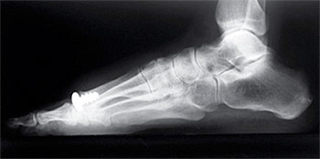
FRANKLIN, Mass.Sept. 12, 2017 /PRNewswire/ — Arthrosurface, Inc. announced today the publication of long-term HemiCAP® data in The Journal of Foot and Ankle Surgery, “10 Year Follow-Up of Metatarsal Head Resurfacing Implants for Treatment of Hallux Rigidus.” The study reported excellent pain relief, functional improvement, high patient satisfaction, a low reoperation rate and no serious complications.

Hallux Rigidus describes a painful condition which affects the big toe at the first metatarsal joint. Osteophytes, or bone spurs, develop on the dorsal surface of the bones involved, which limit motion and cause pain. Advanced disease may also include degenerate cartilage on the joint surfaces, which can contribute to patient pain.

Historically, arthrodesis, or joint fusion, was the standard of care to address patients’ symptoms in advanced stages, however, with eliminated joint motion, patients have shown difficulties with rising on their toes, kneeling or taking a full stride when walking. Other complications include non-union of the fusion site in about 10% of the procedures.

The proprietary HemiCAP® Implant System is designed to maximize implant stability through a strong threaded fixation component and optimizes the fit with a range of implant curvatures. As an active alternative to joint fusion, the HemiCAP® System allows physicians to provide patient specific solutions with hemiarthroplasty of the first and lesser toes, as well as total joint replacement of the big toe. The motion preserving platform provides a complete solution for primary and revision surgery, while maintaining an exit into joint fusion. To date more than 33,000 implants have been used.

“Patients today want to maintain their toe mobility to stay active, whether it is playing golf, performing yoga or being able to continue to live independently,” said Lisa Donnelly VP of Marketing. “We see tremendous traffic on our website from patients inquiring how to find a physician who is trained on our technologies. We have recently launched a new physician locator covering a nationwide network of approximately 1,000 foot and ankle surgeons to help assist them.”

“It is very satisfying to see the AOFAS score of 90.6 at the 10-year follow-up and the high degree of patient satisfaction,” said Steve Ek, President & CEO of Arthrosurface. “We hope this data continues to drive the clinical and patient communities to question the role of motion sacrificing procedures in the first MTP joint, as has happened over time in multiple other joints”.

About Arthrosurface

Arthrosurface, Inc. is a global orthopedic medical technology business providing a broad portfolio of essential products and instrumentation used to treat upper and lower extremity orthopedic conditions caused by trauma, injury and arthritic disease. The product offerings include devices, instruments and orthobiologics designed to preserve and restore the joints so patients can regain and maintain an active lifestyle. The Company offers a variety of unique systems that provide less invasive technologies for surgeons that can be used to treat a wide range of joint conditions. Founded in 2002, Arthrosurface markets and distributes its products in the US and around the world and has succeeded in helping patients return to activity for over 13 years. For more information, please go to our website at www.arthrosurface.com


SOURCE Arthrosurface, Inc.

First Spinal Cord Stimulator Implant Placed in Northern California by Dr. Goldthwaite

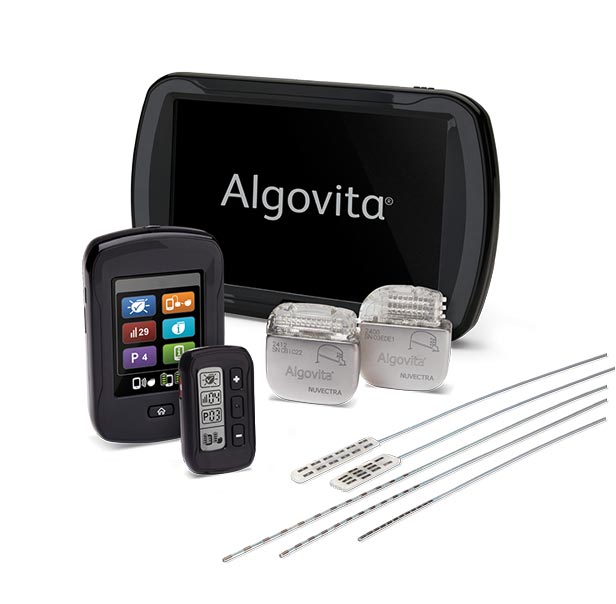
PLANO, Texas, Sept. 11, 2017 (GLOBE NEWSWIRE) — Noel Goldthwaite, MD, senior surgeon and founding member at SpineCare Medical Group, Inc., is the first physician in northern California to implant Nuvectra’s Algovita® Spinal Cord Stimulation System, which successfully treated a local injured veteran’s chronic pain and led to his significant reduction in opioid use. The Nuvectra Algovita SCS System is a powerful and versatile spinal cord stimulation system that includes the smallest patient controller on the market. Algovita is FDA approved as a treatment for chronic, intractable pain.

“Following injuries sustained in the line of duty, this particular patient had multiple surgeries that led to unmanaged and intense chronic back pain prior to the placement of Algovita,” Dr. Goldthwaite explains. “The smaller volume paddle lead and ability to steer it using a stylet eased the navigation of significant scar tissue during the procedure. These features allowed me to place the paddle and stimulation exactly where the patient needed it, which helped to alleviate his pain right from the start. Furthermore, after the procedure, the Algovita system differs from other SCS systems in its easy to use keychain patient controller that allows the patient, in conjunction with our medical staff, to customize the amplitude, frequency, and pulse width of the stimulation and toggle between therapy settings. The results have been outstanding! When he initially came to see me, he was severely depressed and in a wheelchair due to his pain. Now he walks into my office upright with a smile on his face and a sparkle in his eye – he’s a totally different person.”

Chronic pain affects over 100 million Americans according to the Institute of Medicine, more than diabetes, cancer, and heart disease combined. Chronic pain is difficult to treat, but one potential treatment option is spinal cord stimulation, or SCS. SCS works by masking or interrupting pain signals as they travel up the spinal cord to the brain. A small, rechargeable battery is implanted under the patient’s skin and connected to thin wires, called leads, which stimulate the nerves sending pain signals. The stimulation “overrides” the pain signals, and instead of feeling pain, patients may feel a tingling sensation known as paresthesia.

Dr. Goldthwaite also discussed the opioid dependency epidemic occurring among our veterans, stating that, “SCS is a powerful non-drug tool that can be used to control pain relief for these patients. Non-drug pain control helps them function, focus, and sleep better and more effectively deal with other related conditions, including PTSD.”

Algovita gave near immediate pain relief to this patient, enabling him to reduce his opioid usage by over 90% in the 6 months since his surgery, which he had become dependent on to keep the pain at bay. Now, he leads what he calls a life of conscious living, being able to enjoy time with his family and help other veterans who have returned from active duty.

The Algovita SCS System was designed to help physicians improve their outcomes and better manage their patients’ pain today and into the future. Algovita offers a number of unique features not available with other systems on the market. With the broadest range of capabilities and fine-tuned control, Algovita provides physicians with the tools to address the individual and changing nature of pain. In addition, Algovita’s leads cover the longest span of the spinal nerves, allowing for additional coverage options as pain moves and changes. As the only stretchable leads on the market, Algovita’s leads are designed to bend and stretch with body movement to help sustain the therapy long-term. Since pain can change throughout the day and with different activities, Algovita also features a small, wireless, key-fob patient controller so stimulation can be easily and discreetly managed.

For more information about Nuvectra and the Algovita Spinal Cord Stimulation system, visit www.nuvectramed.com.

About Dr. Goldthwaite

Dr. Noel Goldthwaite was a founding member of SpineCare Medical Group in 1988.  He has performed more than 12,000 spinal operations including more than 350 spinal cord stimulator system implantations and revisions both for the lumbar spine and the cervical spine.  He lectures and teaches nationally and internationally.  He has a special interest in treating patients who have already had unsuccessful spine surgeries.  He understands very well that inadequately treated spine problems, especially failed surgeries with chronic pain, are devastating for the patient, the patient’s family, the patient’s friends, and for society.  He is prepared to address all those issues.

Cerapedics Announces Publication of Two-Year IDE Study Follow-Up Data on i-FACTOR™ Bone Graft in the Cervical Spine

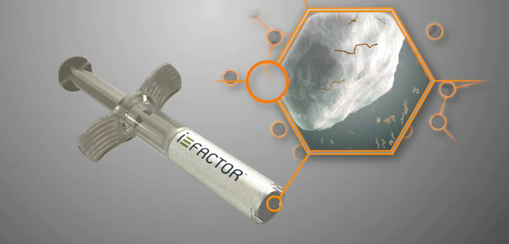
WESTMINSTER, Colo.Sept. 12, 2017 /PRNewswire/ — Cerapedics, a privately-held orthobiologics company, today announced the publication of two-year follow-up data from an FDA Investigational Device Exemption (IDE) clinical trial of i-FACTOR™ Peptide Enhanced Bone Graft. The results published in the peer reviewed journal Neurosurgery show i-FACTOR Bone Graft is statistically superior in overall clinical success to autograft in anterior cervical discectomy and fusion (ACDF) procedures.

i‐FACTOR Bone Graft is based on synthetic small peptide (P-15) technology developed by Cerapedics to support bone growth through cell attachment and activation. In November 2015, Cerapedics received Premarket Approval (PMA) from the FDA for the use of i-FACTOR Bone Graft in ACDF procedures.

“We are pleased that the statistical superiority in overall success for i-FACTOR Bone Graft versus autograft in ACDF procedures was maintained over two years in our IDE trial,” said Jeffrey Marx, Ph.D., president and COO of Cerapedics. “The Neurosurgery publication is a testament to the hard work and dedication of the clinical investigators who continue to pursue advanced treatment options for patients with degenerative cervical disc disease.”

In the IDE clinical trial, patients received either autograft or i-FACTOR Bone Graft in a cortical ring allograft. FDA-mandated success criteria included fusion, improvement in Neck Disability Index (NDI), neurological status, and safety. At two-year follow-up, a responder analysis of combined endpoints for overall success demonstrated 70 percent success for patients receiving i-FACTOR Bone Graft versus 56 percent for patients receiving autograft. This was statistically significant for superiority (p = 0.0302).

Fusion success was confirmed radiologically for 97 percent of i-FACTOR Bone Graft patients and 94 percent of autograft patients (p = 0.2195). A more than 15-point improvement from baseline NDI was reported for 77 percent of i-FACTOR Bone Graft patients and 69 percent of autograft patients (p = 0.1804). Improvement in neurological status was similar in both patient groups (95 percent of i-FACTOR Bone Graft patients and 94 percent of autograft patients, p = 0.6944). Safety success measured by the absence of re-operation, device explantation, and device-related serious adverse events was observed in 95 percent of i-FACTOR Bone Graft patients and 91 percent of autograft patients (p = 0.1379).

“The publication of the two-year results from this Level I clinical trial further validates previous findings that i-FACTOR™ Bone Graft, with P-15 technology, is statistically superior to autograft,” said Paul Arnold, M.D., neurosurgeon at the University of Kansas Hospital Marc A. Asher, M.D. Comprehensive Spine Center and lead author of the study. “we are pleased to be able to offer a new advanced biologic bone graft with outstanding safety and efficacy to our patients.”

About Cerapedics

Cerapedics is an orthobiologics company focused on developing and commercializing its proprietary synthetic small peptide (P-15) technology platform. i-FACTOR Peptide Enhanced Bone Graft is the only biologic bone graft in orthopedics that incorporates a small peptide as an attachment factor to stimulate the natural bone healing process. This novel mechanism of action is designed to support safer and more predictable bone formation compared to commercially available bone growth factors. More information can be found at www.cerapedics.com.

Media contact:
Adam Daley
|Berry & Company Public Relations
212-253-8881
adaley@berrypr.com

SOURCE Cerapedics

Medtronic to set up Israeli R&D centers

11 Sep, 2017 – Yuval Azulai and Gali Weinreb

Medical equipment company Medtronic will be the first company to establish a development center in Israel in cooperation with the Israel Innovation Authority. In return for hiring 100 employees in spheres beyond the company’s existing activity in Israel, the company will receive a $14 million grant from the Innovation Authority. Following approval of the grant in recent days, the new deal between the state and Medtronic is a handshake away.

It is believed that the grant will be spread over three years by paying 30% of the salary of those working in the development center. The new activity will be in two locations: Jerusalem and Yokneam, and will involve brain monitoring (Jerusalem) and big data for medicine (Yokneam).

The expansion of Medtronic’s activity in Israel was agreed four months ago in the framework of a meeting between Ministry of Economy and Industry Eli Cohen, Israel Innovation Authority CEO Aharon Aharon, and Medtronic chairman and CEO Omar Ishrak. A senior source in the Ministry of Economy and Industry said today that in addition to a number of contacts at various stages taking place between the state and multinationals interested in investing in the Israeli economy, initial negotiations were also being conducted with a major pharmaceutical company.

The source said, “The background to these companies’ interest in investments in Israel involving the opening of development or manufacturing centers is the attractive tax environment in comparison with other countries and the low tax rate on intellectual property.”

Other Ministry of Economy and Industry sources said that talks were taking place today with representatives of 10 multinationals for possible investment in Israel. The sources claim that a low media profile is being maintained in these contacts in order to avoid wrecking them.

According to Innovation Authority figures, foreign investment in Israel totaled $12.6 billion in 2016, 7% more than in 2015. The Innovation Authority said that 320 multinationals were operating in Israel.


READ THE REST HERE


What Factors Are Important to Patients When Deciding Between Minimally Invasive or Open Surgery for Posterior Lumbar Fusion?

Written by 

Research on whether minimally invasive surgery (MIS) for lumbar spine fusion is more advantageous than open surgery has been the topic of much debate. While studies suggest less blood loss and fewer infections with MIS, this approach may take longer and be more technically demanding.

Given the lack of consensus on which approach may be best, patient education and shared decision-making can be complex. New findings suggest that patient preference may be influenced by many factors during consent-taking, as reported in the August issue of The Spine Journal.

“Our study demonstrated that patients change their decisions between MIS and open approaches frequently during the process of consent-taking,” said lead author Hwee Weng Dennis Hey, FRCSEd (Orth), FAMS (Orth), MCI, Consultant, Orthopaedic and Spine Surgery, University Orthopaedics, Hand and Reconstructive Microsurgery Cluster, National University Health System in Singapore. “This is likely due to the different weight each patient gives to the advantages and disadvantages of MIS or open surgery.”

“The most common concerns influencing patients’ final decision are radiation and cosmesis,” Dr. Hey told SpineUniverse. “As a result, consent-taking should be conscientious such that patients are adequately informed. Radiation and cosmesis should be given more emphasis.”

Cross Sectional Study of Patients with Lumbar Spondylolisthesis
The findings are based on a cross-sectional study of 54 patients (mean age, 55.8 years) collected over a 3-month period at a single institution in Singapore. These patients were indicated for transforaminal lumbar interbody fusion (TLIF) for radiographically confirmed lumbar spondylolisthesis. All patients had chronic low back pain and radiculopathy. Patients who had undergone previous spine surgery, had concurrent spine pathologies (eg, tumors, infection, or fractures), or had high-grade spondylolisthesis were excluded from the study.

Standardized Consent-Taking Process
An independent spine specialist interviewed patients using a script that required patients to decide between MIS or open surgery at various points during the consent-taking process. Pictures of the scars from MIS and open surgery as well as information on the potential advantages and disadvantages of both approaches were also provided.


READ THE REST HERE

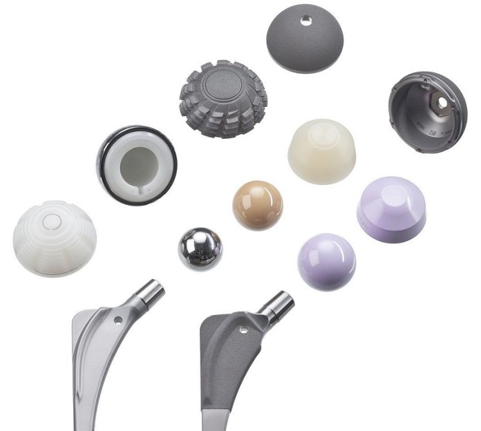
Hip Reconstruction Devices US $5.68 Billion in 2016 and is Expected to Rise $126.6M Per Annum From 2017-2022: Transparency Market Research

ALBANY, New YorkSeptember 8, 2017 /PRNewswire/ —

According to the research report, the global market for Hip Reconstruction Devices stood at US$5.68 bn in 2016 and is expected to witness strong growth over the period from 2017 to 2022. Primary cemented hip replacement devices enjoy a much higher demand among all the products available in this market. With a rise to US$126.6 mn per annum in revenues, this segment is anticipated to retain its dominance over the forecast period. With an increasing patient pool, osteoarthritis and rheumatoid arthritis have emerged as the primary indication for the application of these devices. Researchers predict this trend to continue over the next few years. Hip reconstruction devices find significant application in hospitals, ambulatory surgical centers (ASCs), and orthopedic clinics.

View Full Report at http://www.transparencymarketresearch.com/hip-reconstruction-devices-market.html

“With the significant increase in the geriatric population base across the world, the prevalence of osteoporosis and rheumatoid arthritis is augmenting continuously, resulting in the soaring need for hip reconstruction surgeries, which, subsequently, is boosting the demand for hip reconstruction devices,” says a TMR analyst. The rising cases of hip injuries and the growing demand for minimally invasive procedures is also propelling the global hip reconstruction devices market remarkably.

Going forward, the technological advancements in hip reconstruction devices are likely to drive the growth in this market over the next few years. The upswing in the investments for research and development of highly advanced and minimally invasive hip reconstruction procedures and devices is also projected to have a positive impact on this market in the near future.

High Cost of Devices to Act as Restraint

The worldwide hip reconstruction devices market, although, looks thriving; however, the high cost of hip reconstruction procedures and devices may restrict the market from growing steadily in the coming years. The stringent regulations required for the approval for these devices will also create hindrances in the growth trajectory of this market over the next few years.

Nonetheless, the rising awareness pertaining to the benefits of hip construction procedures in treating patients and enabling them to perform daily activities and live a normal lifestyle is expected normalize the impact of these restraints in the years to come, states the study.

Request to View Sample Report – http://www.transparencymarketresearch.com/sample/sample.php?flag=S&rep_id=10307

Regionally, North America has emerged as the leading market for hip reconstruction devices across the world. Analysts at TMR anticipate this regional market for remain on the top over the forecast period on the grounds of the presence of established hip reconstruction device vendors in a large number. The increasing demand for minimally invasive surgeries is also projected to boost the North America market for hip reconstruction devices in the years to come, states the research report.

With a large pool of manufacturers, the global hip reconstruction devices market is demonstrating a high competitive and fragmented business landscape, finds a new research study by Transparency Market Research (TMR). Innovation and advancements in current offerings is the key trend among the leading players in this market, such as United Orthopedic, Exactech, Corin, Braun Melsungen, DePuy Synthes, Stryker and others. However, over the forthcoming years, a shift in their focus towards strategic alliances, in the form of mergers, acquisitions, and partnerships, can be observed, notes the research study.

Hip Reconstruction Devices Market Report IAvailable @ US$ 5795

About TMR

Transparency Market Research (TMR) is a global market intelligence company providing business information reports and services. The company’s exclusive blend of quantitative forecasting and trend analysis provides forward-looking insight for thousands of decision makers. TMR’s experienced team of analysts, researchers, and consultants use proprietary data sources and various tools and techniques to gather and analyze information.
Contact
Transparency Market Research
State Tower
90 State Street,
Suite 700,
Albany NY – 12207
United States
Tel: +1-518-618-1030
USA – Canada Toll Free: 866-552-3453
Email: sales@transparencymarketresearch.com

Website: http://www.transparencymarketresearch.com 
TMR Blog: http://www.theglobalhealthnews.com/

SOURCE Transparency Market Research

EOS Imaging Reports First Half 2017 Financial Results

September 07, 2017

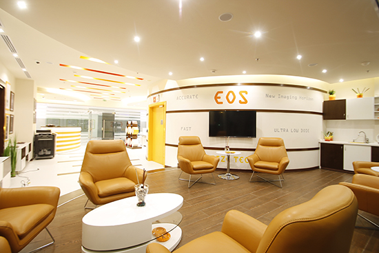
EOS imaging (Paris:EOSI)(Euronext, FR0011191766 – EOSI – Eligible PEA – SME), the pioneer in 2D/3D orthopedic medical imaging, today announced its consolidated results for the six months ended June 30, 2017, as stated by the Board of Directors on September 7, 2017.

First Half 2017 Financial Results

in millions euros First Half 2017 First Half 2016
Sales 16,46 14,14
Other Income 0,82 1,17
Total Income 17,28 15,31
Direct COGS (9,03) (7,63)
Gross Margin

as % of sales

8,25

45,1%

7,68

46,0%

Operating expenses (10,91) (9,41)
Operating Income (2,66) (1,73)
Financial Income (1,06) (0,67)
Net Income (3,72) (2,40)
As of June 30, 2017 As of Dec. 31, 2016
Cash 17,6 14,9

Marie Meynadier, Chief Executive Officer of EOS imaging, commented: “Our performance in the first half of 2017 included strong revenue growth in the EMEA and APAC regions, partially offset by lower sales in North America where we were implementing a new, reinforced organizational structure. This impacted our operational and financial performance, despite the improvement in production and maintenance efficiency.

EOS imaging solutions are increasingly recognised as a standard of care, as evidenced by nearly one million patients each year that benefit from a low-dose EOS scan and comprehensive imaging results, along with the physicians and healthcare systems that benefit from the platform’s innovative and effective solutions. Given this positive position in the market, we decided to accelerate our growth initiatives in the United States, our most important market and a priority for our company.

We are confident in a rapid return to growth in North America, under the leadership of Mike Lobinsky, who we recently appointed as President of the region.

Our initiatives in North America, combined with the positive business dynamics in the rest of the world, and with our investments in our product offering, should strongly improve our performance in the short and medium terms.”

  • First Half 2017 Sales Growth: +16%

In the first half of 2017, the Company generated revenue of €16.5 million, up 16% compared to the first half of 2016. The Company sold 34 EOS® systems during the first half of the year, compared to 28 systems in the same period last year. Sales of maintenance contracts increased by 28% to €2.84 million, reflecting the continued growth of the installed base of EOS systems under contract.

The increase in revenues over the first half of 2017 was driven by strong sales in the EMEA and APAC regions, partially offset by lower sales in North America, related to an operational reorganization.

  • First Half 2017 Gross Margin: €8.3 million representing 45.1% of Revenues

Gross margin for the first half of 2017 was 45.1% of revenues, as compared to 46.0% during the same period last year. This reflects lower sales in North America, which represented approximately 35% of revenues in the period, as compared 54% of revenues in the first half of 2016. This was partially offset by improved manufacturing costs and maintenance in the first half of 2017 compared to the same period last year.

  • Controlled Increase of Operating Expenses

Operating expenses for the first half of 2017 totaled €10.9 million, up 16% compared to the same period last year and in-line with the Company’s revenue growth in the period. These expenses are slightly below their level in the second half of 2016.

Operating loss for the first half of 2017 was €2.7 million, compared with an operating loss of €1.7 million in the same period last year (and of €2.8 million in the second half of 2016). This reflects the decrease in gross margin and a one-time decline in other income (CIR and subsidies).

Net financial expense for the first half of 2017 totaled €1.1 million, compared to €0.7 million in the same period last year, reflecting interest expense on the Company’s €15 million debt financing, the last tranche of which was drawn in June 2016.

Net loss for the first half of 2016 was €3.7 million, compared with a net loss of €2.4 million in the same period last year (and €2.8 million in the second half of 2016).

The Company had 152 employees at June 30, 2017, compared with 132 at December 31, 2016.

  • Cash Position and Balance Sheet: Improving Equity and Cash Positions

The Company’s cash position at June 30, 2017 was €17.6 million, compared with €14.9 million at December 31, 2016. Equity at June 30, 2017 was €27.3 million, compared with €22.8 million at December 31, 2016. This includes 2,204,844 new shares issued primarily in a private placement completed in April 2017 but coming also from the Equity Line Société Générale and from the exercise of options.

  • First Half of 2017 Financial Report

EOS imaging’s first half of 2017 financial report can be downloaded from its web site at www.eos-imaging.com by selecting Documentation, then Financial reports from the Investors menu.

For more information, please visit www.eos-imaging.com.

EOS imaging has been chosen to be included in the new EnterNext© PEA-PME 150 index, composed of 150 French companies and listed on Euronext markets in Paris.

EOS imaging is listed on Compartment C of Euronext Paris

ISIN: FR0011191766 – Ticker: EOSI

About EOS imaging

EOS imaging designs, develops, and markets EOS®, an innovative medical imaging system dedicated to osteo-articular pathologies and orthopaedics, as well as associated solutions. The Company is authorized to market in 51 countries, including the United States (FDA), Japan and the European Union (EC). The Group posted 2016 revenues of €30.8 million and employed 132 people at December 2016, including an R&D team of 43 engineers. The Group is based in Paris and has five subsidiaries in Besançon (France), Cambridge (Massachusetts), Montreal (Canada), Frankfurt (Germany) and Singapore.

Contacts

EOS imaging
Pierre Schwich, CFO
Ph: +33 (0)1 55 25 60 60
investors@eos-imaging.com
or
NewCap
Financial communication and investor relations
Pierre Laurent
Ph: +33 (0)1 44 71 94 96
eosimaging@newcap.eu
or
The Ruth Group (US)
Press relations / Joanna Zimmerman
Ph: 646-536-7006
jzimmerman@theruthgroup.com

Spinal Resources, Inc.®’s Swedge Pedicle Screw System Receives FDA 510(k) Clearance

FT. LAUDERDALE, Fla.Sept. 7, 2017 /PRNewswire/ —  Spinal Resources, Inc.® (SRI) announced that it has received FDA 510(k) clearance for its Swedge™ Pedicle Screw System. This unique system offers multiple benefits, including CoCr and Titanium tulips; double lead and Cortical Cancellous Threads; Polyaxial, Monoaxial, Reduction, and Iliac screws; and MIS (minimally invasive spinal surgery) options. Currently, the Swedge system is cleared for a 5.5mm rod; 4.75mm and 5.0mm rods are pending clearance.

Since 2005, Spinal Resources, Inc. has been developing its groundbreaking technology, which accommodates multiple rods in a single-screw tulip with single-locking cap, and effectively locks the tulip’s angle without the presence of a rod. The FDA’s clearance of the Swedge™ Stabilization System marks the transition of Spinal Resources, Inc. from design and distribution experts into full manufacturers of innovative surgical enhancements. The Swedge system is the company’s first product development effort–and a seminal achievement: SRI’s system holds the tulip’s position while contouring rods in a multi-level scenario. The transition rod features a gradual taper between sections to minimize stress risers. Designed to improve efficiencies within the current and trending hospital environment by reducing inventory and increasing surgical procedure options, which may result in more affordable alternatives for patients, Spinal Resources, Inc.’s versatile, all-in-one Swedge implant system gives clinicians more surgical flexibility in degenerative, deformity, and midline approach surgeries, with a wider range of options to stabilize and conform to varying anatomies. Its MIS capabilities offer patients a smaller incision, with the added benefits of less tissue disturbance, reduced rehab/recovery time, and enhanced outcomes.

A report from GlobalData Healthcare, a UK-based company specializing in providing business data and analytics, indicated the 2017 global spinal fusion market is experiencing steady growth of about 3.4% CAGR (compound annual growth rate). The market is estimated to rise from $7.1 billion in 2016 to approximately $9 billion by 2023. The December 2016 report attributes the increase to degenerative spinal conditions caused by aging and the prevalence of technological advancements in surgeries. To learn more, see https://www.globaldata.com/spinal-fusion-market-approach-9-billion-2023-technology-improves-says-globaldata/.

According to President and Chief Executive Officer Bernard Bedor, no other screw on the market is quite like the Swedge System in capability. “We believe we’ve done something unique with our product,” he said. “We’ve taken three different rod sizes, put them into one effective tulip, and can lock them with a one-step locking cap. The Swedge System provides surgeons with greater versatility in the pre-planning and inter-operative stages, effectively reducing the surgical ‘fiddle factor’ thanks to its convenient standard and transition rods and screws in a variety of diameters and lengths. Our focus at SRI is to be problem-solvers for patients by supporting surgeons and hospitals in their important work with fewer instruments and a smarter system that shorten operating room time and lower costs.”

With the ownership of four (4) patents (and two more under consideration), this FDA clearance, and several other promising products in its pipeline, SRI is standing out as a strong contender in the design and manufacture of innovative solutions that enhance surgical procedures. The company is exploring full commercialization via strategic partnership, license, and/or private/corporate funding. For more information, contact our media and investors relations specialist: Ms. Melanie Raskin – melanie.raskin@aktaPD.com

Intended Use
The Swedge™ Pedicle Screw Fixation System is intended to provide immobilization and stabilization of spinal segments in skeletally-mature patients as an adjunct to fusion in the treatment of acute and chronic instabilities or deformities of thoracic, lumbar, and  sacral spine: degenerative spondylolisthesis with objective evidence of neurological impairment, fracture, dislocation, deformities or curvatures (i.e., scoliosis, kyphosis, and/or  lordosis), spinal tumor, pseudoarthrosis, and failed previous fusion.

It is also intended for non-cervical pedicle screw fixation for the following indications: severe spondylolisthesis (grades 3 and 4) of the L5-S1 vertebra in skeletally-mature patients receiving fusion by autogenous bone graft, having implants attached to the lumbar and sacral spine (L3 to sacrum) with removal of the implants after the attainment of a solid fusion. It is also intended for the following indications: DDD (degenerative disc disease), trauma (i.e., fracture or dislocation), spinal stenosis, deformities or curvatures (i.e., scoliosis, kyphosis, and/or lordosis), tumor, pseudoarthrosis, and failed previous fusion.

About Spinal Resources, Inc.® (SRI)
Founded in 2005, Spinal Resources, Inc.® is a Ft. Lauderdale, Florida-based spinal medical device company that supports cost-effective patient care with innovative mechanical and biomechanical products to alleviate pain, shorten recovery time, restore health, and extend quality of life. SRI has engineered a unique line of patented spinal and cervical fixation systems with stem growth factors for the surgical repair and reconstruction of debilitating spinal conditions. For more information, visit spinalresourcesinc.com.

Media Contact:
Melanie Raskin
175102@email4pr.com


SOURCE Spinal Resources, Inc.(R)