Trice Medical, Inc. and Pacific Medical, Inc. announces multi-state distribution agreement.

2/2/2017

King of Prussia, PA., and Tracy, CA.. – reported today on the completion of a distribution agreement with Pacific Medical, Inc., a distributor of durable medical equipment in the United States who specializes in orthopedic, rehabilitation, surgery and sports medicine.

Pacific Medical will be responsible for the promotion and distribution of the mi-eye 2™, an in-office arthroscopic diagnostic tool, in Northern California, Northern Nevada and the Pacific Northwest including Oregon, Washington, Alaska, Idaho and Montana. The partnership with Pacific Medical puts the mi-eye 2 in the hands of 300 dedicated sales and service personnel. Pacific Medical has long been a pioneer of surgeon and physician training, including a 12-station state of the art wet bioskills lab, which includes a 300-seat auditorium that Trice Medical and PacMed will utilize to enhance surgeon education throughout the Pacific Northwest.

“Trice Medical is thrilled to have PacMed as our distribution partner for the mi-eye 2 in the Northwest. The PacMed team has a long history of educating Orthopedic Surgeons on new technology. They are focused on partnering with their customers to deliver value to physicians, payers and patients.” Said Mark Foster, Chief Commercialization Officer for Trice Medical. “The mi-eye 2 is designed to deliver INSTANT ANSWERS to those same stakeholders and we are looking forward to successfully expanding our reach into the Northwest.”

Paul Wiese, VP of Sales for Pacific Medical stated. “The mi-eye 2 will allow us to continue in our mission of providing innovative technologies and advanced modalities that help our surgeon partnerships in their pursuit of increasing the surgeon-patient experience. Surgeons will be able to confirm their clinical diagnosis same day and eliminate the potential need for patients to take addition time away from work or leisure, to get the answers to their injuries.  The mi-eye 2 technology allows the physician to immediately plan the care of their patient, can take the guesswork out of MRI’s, and patients will now be intimately involved with their surgeons in the care of their injuries. We are very excited about our partnership with Trice Medical and the impact this technology will have on improving the quality of healthcare services provided by our physicians.”

About Trice Medical, Inc. 

Trice Medical was founded to fundamentally improve orthopedic diagnostics for the patient, physician, and payor by providing instant answers. Trice Medical has pioneered fully integrated camera-enabled technology, the mi-eye 2, that provides a clinical solution optimized for the physician’s office. Trice Medical’s mission is to provide more immediate and definitive patient care, eliminating the false reads associated with current indirect modalities and significantly reduce the overall cost to the healthcare system.

Trice Medical is a venture capital backed company; Safeguard Scientifics

(NYSE:SFE), BioStar Ventures and HealthQuest Capital are its major investors; smaller firms and angel investors have completed the rounds.

For more information, visit tricemedical.com or call 844.643.9300.

About Pacific Medical, Inc.

Pacific Medical, Inc. is a distributor of durable medical equipment, sports medicine medical devices, and orthotics; specializing in orthopaedic rehabilitation, arthroscopic surgery, and sports medicine.  Our corporate headquarters are in Tracy, California. Pacific Medical operates in seven western states, with a sales force of more than 100 highly skilled representatives. Incorporated in 1992, the Company now represents several industry leading manufacturers in the states of California, Nevada, Oregon, Washington, Montana, Alaska, and Idaho. This exponential growth has been very carefully planned and charted by an experienced management team.  Our success in the volatile healthcare industry can be directly attributed to the strong relationships forged with our customer base of orthopaedic surgeons, hospital administrators, physical therapists, and trainers.


https://www.pacmedical.com/

BACK


Vericel Announces First MACI Implant in the United States for the Treatment of Symptomatic Cartilage Defects of the Knee

CAMBRIDGE, Mass., Feb. 01, 2017 (GLOBE NEWSWIRE) — Vericel Corporation (NASDAQ:VCEL), a leading developer of expanded autologous cell therapies for the treatment of patients with serious diseases and conditions, today announced the first implant of MACI®(autologous cultured chondrocytes on porcine collagen membrane) in the United States for the repair of symptomatic single or multiple full-thickness cartilage defects of the knee with or without bone involvement in adults.

“While surgeons have long understood that autologous chondrocyte implantation can repair cartilage tissue, the previous surgical procedure was more invasive, technically demanding, time consuming and could result in an uneven distribution of cells,” said Dr. David Recker, chief medical officer of Vericel.  “With the introduction of MACI, surgeons now have an FDA-approved product in which the patient’s own cells can be reproducibly delivered using less invasive techniques.  The MACI implant not only ensures more uniform distribution of the cells in the cartilage defect, but also greatly simplifies the implantation procedure.”

“Treating the first patient with MACI this soon following FDA approval is incredibly rewarding for everyone at Vericel,” said Nick Colangelo, president and CEO of Vericel.  “We believe that the simplified MACI surgical technique, together with the MACI Phase 3 clinical data demonstrating statistically significantly greater improvement in pain and function scores compared to microfracture, will drive significant growth for our cartilage repair franchise.  We are now focused on realizing this growth potential by providing training to expand the number of implanting surgeons and working with payers to ensure reimbursement for MACI.”

About MACI
MACI® (autologous cultured chondrocytes on porcine collagen membrane) is an autologous cellular scaffold product that is indicated for the repair of symptomatic single or multiple full-thickness cartilage defects of the knee with or without bone involvement in adults.  The MACI implant consists of autologous cultured chondrocytes seeded onto a resorbable Type I/III collagen membrane.  Autologous cultured chondrocytes are human-derived cells which are obtained from the patient’s own cartilage for the manufacture of MACI.

The FDA approval of MACI was supported by the results of SUMMIT trial1, a Phase 3 two‑year, prospective, multicenter, randomized, open-label, parallel-group study that enrolled a total of 144 patients, ages 18 to 54 years, with at least one symptomatic Outerbridge Grade III or IV focal cartilage defect on the medial femoral condyle, lateral femoral condyle, and/or the trochlea.  The co-primary efficacy endpoint was change from baseline to Week 104 for the subject’s Knee injury and Osteoarthritis Outcome Score (KOOS) in 2 subscales:  Pain and Function (Sports and Recreational Activities [SRA]).2  At Week 104, KOOS pain and function (SRA) had improved from baseline in both treatment groups, but the improvement was statistically significantly (p<0.001) greater in the MACI group compared with the microfracture group.  In a responder analysis, the proportion of subjects with at least a 10‑point improvement in both KOOS pain and function (SRA) was greater in the MACI group (63/72 = 87.5%; 95% CI [77.6%, 94.6%]) compared with the microfracture group (49/72 = 68.1%; 95% CI [56.0%, 78.6%]).

The most frequently occurring adverse reactions (≥5%) reported for MACI in the 2‑year randomized, controlled clinical trial were arthralgia, tendonitis, back pain, joint swelling, and joint effusion.  Serious adverse reactions reported for MACI were arthralgia, cartilage injury, meniscus injury, treatment failure, and osteoarthritis.

Important Safety Information

  • MACI is contraindicated in patients with a known history of hypersensitivity to gentamicin, other aminoglycosides, or products of porcine or bovine origin.  MACI is also contraindicated for patients with severe osteoarthritis of the knee, inflammatory arthritis, inflammatory joint disease, or uncorrected congenital blood coagulation disorders.  MACI is also not indicated for use in patients who have undergone prior knee surgery in the past six months, excluding surgery to procure a biopsy or a concomitant procedure to prepare the knee for a MACI implant.
  • MACI is contraindicated in patients who are unable to follow a physician-prescribed post-surgical rehabilitation program.
  • The safety of MACI in patients with malignancy in the area of cartilage biopsy or implant is unknown.  Expansion of present malignant or dysplastic cells during the culturing process or implantation is possible.
  • Patients undergoing procedures associated with MACI are not routinely tested for transmissible infectious diseases.  A cartilage biopsy and MACI implant may carry the risk of transmitting infectious diseases to healthcare providers handling the tissue.  Universal precautions should be employed when handling the biopsy samples and the MACI product.
  • Final sterility test results are not available at the time of shipping.  In the case of positive sterility results, health care provider(s) will be contacted.
  • To create a favorable environment for healing, concomitant pathologies that include meniscal pathology, cruciate ligament instability and joint misalignment, must be addressed prior to or concurrent with the implantation of MACI.
  • Local treatment guidelines regarding the use of thromboprophylaxis and antibiotic prophylaxis around orthopaedic surgery should be followed.  Use in patients with local inflammations or active infections in the bone, joint, and surrounding soft tissue should be temporarily deferred until documented recovery.
  • The MACI implant is not recommended during pregnancy.  For implantations post-pregnancy, the safety of breast feeding to infant has not been determined.
  • Use of MACI in pediatric patients or patients over 55 years of age has not been assessed.
  • The most frequently occurring adverse reactions reported for MACI (≥5%) were arthralgia, tendonitis, back pain, joint swelling, and joint effusion.
  • Serious adverse reactions reported for MACI were arthralgia, cartilage injury, meniscus injury, treatment failure, and osteoarthritis.

About Articular Cartilage Defects of the Knee
Articular cartilage is a highly organized avascular tissue composed of chondrocytes embedded within an extracellular matrix of collagens, proteoglycans and noncollagenous proteins.  Its primary function is to enable the smooth articulation of joint surfaces, and to cushion compressive, tensile and shearing forces.  Articular cartilage damage is caused by both acute and repetitive trauma resulting in knee pain, effusion or mechanical symptoms such as catching and locking, and swelling.  Since articular cartilage is avascular it has little capacity to repair itself or regenerate.  Articular cartilage lesions that are left untreated may progress to debilitating joint pain, dysfunction, and osteoarthritis.3  The prevalence rate for cartilage lesions in the knee has been reported to be 63% in patients undergoing investigational arthroscopies.4

About Vericel Corporation
Vericel develops, manufactures, and markets expanded autologous cell therapies for the treatment of patients with serious diseases and conditions.  The company currently markets two cell therapy products in the United States.  Carticel® (autologous cultured chondrocytes) is an autologous chondrocyte implant for the treatment of cartilage defects in the knee in patients who have had an inadequate response to a prior arthroscopic or other surgical repair procedure.  Epicel® (cultured epidermal autografts) is a permanent skin replacement for the treatment of patients with deep dermal or full thickness burns greater than or equal to 30% of total body surface area.  Vericel is marketing MACI® (autologous cultured chondrocytes on porcine collagen membrane), an autologous cellularized scaffold product indicated for the repair of symptomatic, single or multiple full-thickness cartilage defects of the knee with or without bone involvement in adults, which has just been approved by the FDA.  Vericel is also developing ixmyelocel‑T, an autologous multicellular therapy intended to treat advanced heart failure due to ischemic dilated cardiomyopathy (DCM).  For more information, please visit the company’s website at www.vcel.com.

Epicel®, Carticel®, and MACI® are registered trademarks of Vericel Corporation. © 2017 Vericel Corporation. All rights reserved.

This document contains forward-looking statements, including, without limitation, statements concerning anticipated progress, objectives and expectations regarding the commercial potential of MACI® and our other products, and timing, and objectives and expectations regarding our company described herein, all of which involve certain risks and uncertainties. These statements are often, but are not always, made through the use of words or phrases such as “anticipates,” “intends,” “estimates,” “plans,” “expects,” “we believe,” “we intend,” and similar words or phrases, or future or conditional verbs such as “will,” “would,” “should,” “potential,” “can continue,” “could,” “may,” or similar expressions. Actual results may differ significantly from the expectations contained in the forward-looking statements. Among the factors that may result in differences are the inherent uncertainties associated with competitive developments, clinical trial and product development activities, regulatory approval requirements, estimating the commercial potential of our products and product candidates, market demand for our products, product performance and our ability to supply or meet customer demand for our products. These and other significant factors are discussed in greater detail in Vericel’s Annual Report on Form 10-K for the year ended December 31, 2015, filed with the Securities and Exchange Commission (“SEC”) on March 14, 2016, Quarterly Reports on Form 10-Q and other filings with the SEC. These forward-looking statements reflect management’s current views and Vericel does not undertake to update any of these forward-looking statements to reflect a change in its views or events or circumstances that occur after the date of this release except as required by law.

References

1Saris D, Price A, Widuchowski W, Bertrand-Marchand M, Caron J, Drogset JO, et al. Matrix-applied characterized autologous cultured chondrocytes versus microfracture: two-year follow-up of a prospective randomized trial. Am J Sports Med. 2014 Jun;42(6):1384-94.

2Roos EM, Lohmander LS. The Knee injury and Osteoarthritis Outcome Score (KOOS): from joint injury to osteoarthritis. Health Qual Life Outcomes. 2003;1:64.

3Bedi A, Feeley BT, Williams RJ. Management of articular cartilage defects of the knee. J Bone Joint Surg Am. 2010;92(4):994‑1009.

4Curl WW, Krome J, Gordon ES, Rushing J, Smith BP, Poehling GG. Cartilage injuries: a review of 31,516 knee arthroscopies. Arthroscopy. 1997;13(4):456-60.

CONTACT:
Chad Rubin
The Trout Group crubin@troutgroup.com 
(646) 378-2947
or
Lee Stern
The Trout Group lstern@troutgroup.com
(646) 378-2922

Merete was issued New US Patent for Lapidus plantar fixation

January 31, 2017

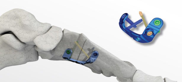
Merete was granted US Patent No. 9,545,276, covering a Lapidus plantar fixation device. The Metafix PlantarMax plate is designed as the first plantar/medial Lapidus fixation plate which takes advantage of the biomechanically superior fixation on the tension side of the MTC (Metatarsal/Cuneiform) joint during a Lapidus Hallux Valgus correction procedure. A medial incision allows adequate exposure of the MTC joint for both joint preparation and plate fixation. As cited in literature, fixation on the tension side of the joint is superior to dorsal or medial fixation and offers additional compression as the patient proceeds to weight bearing. The MetaFix PlantarMax Lapidus Plate is designed with 2 plantar locking screws, an interfragmentary screw hole and 2 medial locking (or non locking) screws for additional fixation.

For further information please contact: Jeff Johnson, National Sales and Marketing Manager

Merete Technologies, Inc., One Lincoln Center, 18W140 Butterfield Rd., 15th Floor, Oakbrook Terrace, IL 60181

Phone: 630‐613‐7181, Fax: 630‐613‐7184, service@merete‐medical.com

Paragon 28 announces launch of unprecedented small bone fixation system for foot & ankle-Baby Gorilla Mini Plating System

ENGLEWOOD, Colo., Jan. 30, 2017 /PRNewswire/ — Since its inception, Paragon 28 has obsessed over every aspect of foot and ankle surgery. Committed to creating tailored solutions to improve surgical outcomes, Paragon 28 has launched innovative products and instrumentation that help to streamline procedures, allow surgeons flexibility in technique and approach, and facilitate reproducible results benefitting both the surgeon and patient.

Today, Paragon 28 continues to honor a tradition of comprehensive and innovative solutions with the launch and release of the Baby Gorilla® Mini Plating System.

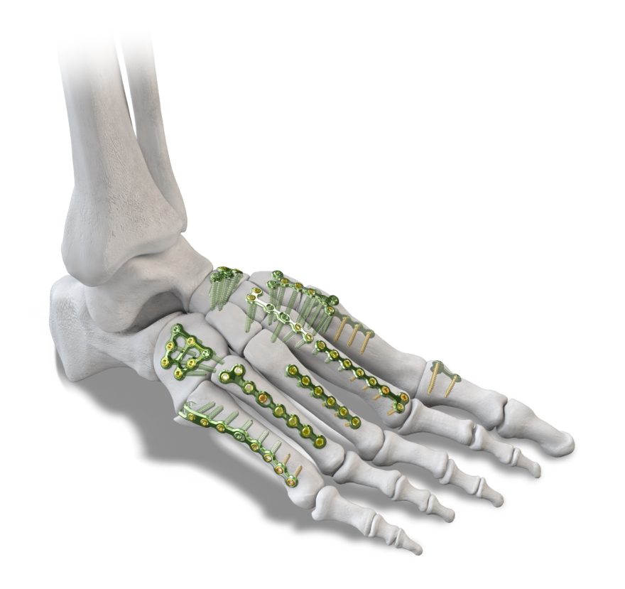
The Baby Gorilla® Mini Plating System leverages the modularity, optionality and specificity admired in the classic Gorilla® Plating system and offers a comprehensive set of implants for procedures that may require narrower plates and smaller screws matching the size and anatomy of small bones within the foot and ankle.

The system provides 65 unique Baby Gorilla® plating options from 13 plating families.  All plates are designed to accept both 2.0 mm and 2.5 mm locking and non-locking variable angle plate screws.

The system offers the most extensive array of universal utility, fracture, and procedure specific plates which require minimal manipulation to achieve satisfactory results.

Plate options found within the Baby Gorilla® Plating System include:

The Baby Gorilla® Mini Plating System also includes a robust assortment of specially designed instrumentation including curettes, reduction clamps, osteotomes, plate bending tools and compression and distraction devices engineered specifically for use in the foot and ankle for small bone fractures and reconstruction procedures.

Paragon 28 is grateful for the significant contributions Dr. Patrick Yoon, MD Hennepin County Medical Center Minneapolis, MN and Dr. Thomas Harris, MD UCLA-Harbor Medical Center, Torrance, CA made as surgeon designers of this system.

About Paragon 28, Inc.

Paragon 28, Inc. was established in 2010 to address the unmet and under-served needs of the foot and ankle community. From the onset, Paragon 28 has made it our goal to re-invent the space of foot and ankle surgery. We believe that through research and innovation we can create new and improved solutions to the challenges faced by foot and ankle specialists.

Product Page:
http://www.paragon28.com/by-system/baby-gorilla/


SOURCE Paragon 28, Inc.

$7.82 Billion Foot and Ankle Devices (Prostheses, Bracing and Support Devices and Orthopedic Implants and Devices) Market 2016 – Forecast to 2025

Dublin, Jan. 26, 2017 (GLOBE NEWSWIRE) — Research and Markets has announced the addition of the “Foot and Ankle Devices Market Analysis & Trends – Product (Prostheses, Bracing and Support Devices and Orthopedic Implants and Devices ), Cause of Injury (Neurological Disorders, Trauma and Diabetes) – Forecast to 2025” report to their offering.

The Global Foot and Ankle Devices Market is poised to grow at a CAGR of around 7.9% over the next decade to reach approximately $7.82 billion by 2025.

Some of the prominent trends that the market is witnessing include increasing numerous sports injuries, growing geriatric population worldwide and increasing new product launches.

Based on product the market is categorized into prostheses, bracing and support devices and orthopedic implants and devices. Prostheses segment is further segmented into Microprocessor-Controlled (MPC) prostheses, Solid Ankle Cushion Heel (SACH) foot, multiaxial prostheses, single-axial prostheses, dynamic response/energy-storing prostheses. Bracing and support devices are further segregated into hinged braces and soft bracing. Orthopedic implants and devices segment is further classified into soft-tissue orthopedic devices, joint implants and fixation devices.

Depending on the cause of injury the market is segmented by neurological disorders, trauma, diabetes and other causes.

This industry report analyzes the global markets for Foot and Ankle Devices across all the given segments on global as well as regional levels presented in the research scope. The study provides historical market data for 2013, 2014 revenue estimations are presented for 2015 and forecasts from 2016 till 2025.

The study focuses on market trends, leading players, supply chain trends, technological innovations, key developments, and future strategies. With comprehensive market assessment across the major geographies such as North America, Europe, Asia Pacific, Middle East, Latin America and Rest of the world the report is a valuable asset for the existing players, new entrants and the future investors.

Key Topics Covered:

1 Market Outline

2 Executive Summary

3 Market Overview
3.1 Current Trends
3.1.1 Increasing Numerous Sports Injuries
3.1.2 Growing Geriatric Population Worldwide
3.1.3 Increasing New Product Launches
3.1.4 Growth Opportunities/Investment Opportunities
3.2 Drivers
3.3 Constraints
3.4 Industry Attractiveness

4 Foot and Ankle Devices Market, By Product
4.1 Prostheses
4.1.1.1 Microprocessor-Controlled (MPC) Prostheses
4.1.1.2 Solid Ankle Cushion Heel (SACH) Foot
4.1.1.3 Multiaxial Prostheses
4.1.1.4 Single-Axial Prostheses
4.1.1.5 Dynamic Response/Energy-Storing Prostheses
4.2 Bracing and Support Devices
4.2.1.1 Hinged Braces
4.2.1.2 Soft Bracing
4.3 Orthopedic Implants and Devices
4.3.1.1 Soft-Tissue Orthopedic Devices
4.3.1.1.1.1 Musculoskeletal Reinforcement Devices
4.3.1.1.1.2 Artificial Tendons & Ligaments
4.3.1.2 Joint Implants
4.3.1.2.1.1 Phalangeal Implants
4.3.1.2.1.2 Ankle Implants (Ankle Replacement Devices)
4.3.1.2.1.3 Subtalar Joint Implants/Subtalar Joint Reconstruction Devices
4.3.1.3 Fixation Devices
4.3.1.3.1.1 Internal Fixation Devices
4.3.1.3.1.1.1.1 Fusion Nails
4.3.1.3.1.1.1.2 Screws
4.3.1.3.1.1.1.3 Plates
4.3.1.3.1.1.1.4 Wires & Pins
4.3.1.3.1.2 External Fixation Devices
4.3.1.3.1.2.1.1 Ring Ankle Fixators
4.3.1.3.1.2.1.2 Unilateral Fixators
4.3.1.3.1.2.1.3 Hybrid Fixators

5 Foot and Ankle Devices Market, By Cause of Injury
5.1 Neurological Disorders
5.2 Trauma
5.3 Diabetes
5.4 Other Causes

6 Foot and Ankle Devices Market, By Geography

7 Leading Companies
7.1 Acumed, LLC
7.2 Arthrex, Inc.
7.3 Extremity Medical, LLC
7.4 Integra Lifesciences Holdings Corporation
7.5 Ossur
7.6 Smith & Nephew PLC.
7.7 Stryker Corporation
7.8 Tornier N.V.
7.9 Wright Medical Technology, Inc.
7.10 Zimmer Biomet Holdings, Inc.
7.11 De Puy Synthes
7.12 Biomet, Inc.
7.13 BioPro, Inc.
7.14 Orthofix Holdings Inc.
7.15 Mondeal Medical Systems GmbH

For more information about this report visit http://www.researchandmarkets.com/research/kphvcg/foot_and_ankle

OIC Launches Intramedullary Tibial Nail

RENO, Nev., Jan. 24, 2017 /PRNewswire/ — The Orthopaedic Implant Company (OIC), a medical device company that specializes in high-value orthopaedic implants, announced the launch of its Tibial Intramedullary Nailing System today.

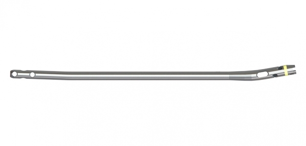
The OIC Tibial Nail System is designed to stabilize fractures of the tibia. Strategically placed proximal and distal screw holes accommodate varying fracture patterns with a proximally-placed slotted hole that allows dynamization.  The nail is available in diameters of 9mm through 11.5mm and lengths of 280mm to 420mm and features 5.0mm diameter cross-lock screws for optimal implant fixation.

The OIC Tibial Nail is part of OIC’s high value IM Nail System and uses the same instrumentation as all IM nails from OIC – decreasing inventory burdens and costs while increasing intra-operative efficiencies.  It is the third nail released in the last twelve months from the company that address long bone fractures. “We are very proud to offer the addition of the OIC Tibial Nail to our high value implant portfolio,” said Todd Martens, VP of Product Design and Customer Experience. “We’ve designed the entire IM Nail System to meet the demands of value based healthcare; a rapidly growing trend.”


About Orthopaedic Implant Company:

OIC entered the medical device market in 2010 with an innovative approach to delivering the highest quality implants at the lowest possible prices. OIC implants cost 50 to 60 percent less than the average market price of premium implants, saving health care systems millions of dollars a year. OIC’s high-quality, low-cost implants and products can be used for a variety of procedures, including treatment of orthopaedic trauma and spinal injuries. 

SOURCE The Orthopaedic Implant Company

Bioness StimRouter™ Neuromodulation System Clinical Data Presented At The 2017 North American Neuromodulation Society’s Annual Meeting

VALENCIA, Calif., Jan. 20, 2017 /PRNewswire/ — Bioness, Inc., the leading provider of state-of-the-art, clinically supported rehabilitation and pain management medical devices, is pleased to announce data surrounding the clinical benefits of its StimRouter Neuromodulation System. At the 2017 North American Neuromodulation Society’s (NANS) Annual Meeting in Las Vegas, Nevada, Dr. Porter McRoberts, a trained Physiatrist and Interventional Spine and Pain Management Specialist in Fort Lauderdale, Florida, shared data indicating that the StimRouter is a promising treatment for post-stroke shoulder pain (PSSP) patients when compared to the alternative options for pain management.

“Post-stroke shoulder pain is a condition that occurs in approximately 30 to 70 percent of stroke patients. This condition contributes to a loss of upper limb use and results in an inability to perform basic daily tasks such as getting dressed or tooth brushing,” said Dr. McRoberts. “While treatments such as oral medication and injections can have a short term impact on managing pain, neuromodulation devices, such as the StimRouter, are designed to target pain at the point of origin and provide relief in a minimally invasive manner with long term effect.”

In a poster presentation, Dr. McRoberts shared data from seven patients that were implanted with the StimRouter in an effort to manage their post-stroke shoulder pain. Using the Visual Analogue Scale (VAS) patients measured pain both before the procedure and up to four months after implant. The patients received care across four different U.S. hospitals in New York, Florida, and Pennsylvania.

Dr. McRoberts and his colleagues found that:

  • PSSP patients implanted with the StimRouter Peripheral Nerve Stimulator experienced an average of 70% reduction in their chronic pain using the Visual Analog Scale (VAS).
  • Peripheral Nerve Stimulation is a promising treatment for PSSP, especially in light of the alternatives for pain management.

The results also show that peripheral nerve stimulation targeting the axillary nerve is a promising treatment for post-stroke shoulder pain patients. Implantable peripheral neurostimulation therapies, like the StimRouter, can be safe and effective pain management techniques as an adjunct to other therapies like Physical/Occupational therapy to help with improving mobility and quality of life.

“The StimRouter has already shown promising results in treating chronic peripheral nerve pain and we are honored to now be sharing its potential for relieving the pain many post-stroke shoulder patients encounter,” said Todd Cushman, President and CEO at Bioness. “Our goal at Bioness is to help patients return to their pre-stroke lives by reducing pain and getting them back into rehabilitation therapy by a Physical/Occupational Therapist. This data is another step forward showing that it is possible for post-stroke patients to regain control of their lives.”

The StimRouter was the first FDA cleared, long-term, minimally invasive peripheral nerve stimulator indicated to treat chronic pain of a peripheral nerve origin. This non-drug, patient-controlled medical device is an adjunct to other modes of therapy and is being well received by patients and clinicians alike.

For more information on the StimRouter or to speak with a patient currently using the device, stop by Bioness’ booth #221 at NANS or visit www.stimrouter.com for videos of real patients sharing their StimRouter experience.

About StimRouter™ Neuromodulation System
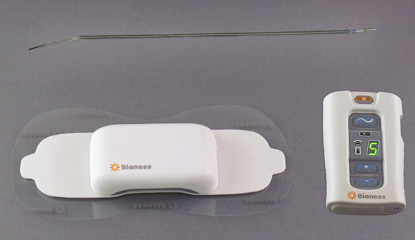
The StimRouter™ Neuromodulation System is intended to provide electrical stimulation via a small implanted lead to a target peripheral nerve, for the management of severe intractable chronic pain of peripheral nerve origin in adults, as an adjunct to other modes of therapy. StimRouter is a minimally invasive procedure consisting of an implanted lead, external pulse transmitter (EPT) and conductive electrode, controlled by a small, handheld wireless patient programmer. StimRouter is programmed at the direction of the physician to meet unique pain management requirements.

About Bioness, Inc. Bioness is the leading provider of innovative technologies helping people control pain, regain mobility and improve independence. Bioness solutions include external and implantable functional electrical stimulation (FES) systems, robotic systems and software-based therapy programs providing functional and therapeutic benefits for individuals affected by pain, central nervous system disorders and orthopedic injuries. Individual results vary. Consult with a qualified physician to determine if this product is right for you. Contraindications, adverse reactions and precautions are available online at www.bioness.com.

Media Relations Contact Information
Next Step Communications
bioness@nextstepcomms.com
781.326.1741

StimRouter™ and Bioness® are trademarks of Bioness, Inc. | www.bioness.com | Rx Only
Additional information about StimRouter can be found at www.stimrouter.com.

SOURCE Bioness, Inc.


OrthoArizona – Arizona Hand, Wrist and Elbow Surgeons Expands its Practice by Adding a New Surgeon to Its Practice

PHOENIX, ARIZONA (PRWEB) DECEMBER 20, 2016

The premier hand, wrist and elbow surgery practice in Phoenix, OrthoArizona – Arizona Hand, Wrist and Elbow Surgeons, is expanding its practice with the addition of fellowship-trained hand surgeon.

Ryan A. Odgers, MD brings with him extensive wrist, microvascular and hand surgery experience. He joins the board-certified team at OrthoArizona – Arizona Hand, Wrist and Elbow Surgeons which also includes E. Scott Frankel, MD; Mark J. Leber, MD; Matthew M. Conklin, M.D.; and Richard K. Hinkle, PA-C.

“We’re confident that Dr. Odgers will continue the level of excellence we’ve established and are excited that he has joined our team,” said Dr. Mark Leber.

Dr. Odgers assumes his new position at Arizona Hand, Wrist and Elbow Surgeons following fellowship training focused on hand and upper extremity surgery at Integris Baptist Medical Center in Oklahoma City. Prior to his fellowship, Dr. Odgers received a bachelor’s degree in biology from Arizona State University, a medical degree from the University of Oklahoma and completed his residency right here in Phoenix.

For more information about OrthoArizona – Arizona Hand, Wrist and Elbow Surgeons, please call (602) 393-1010 or visit http://azhandwristspecialists.com/. Find out more about OrthoArizona – Arizona Hand, Wrist and Elbow Surgeons on Facebook at https://www.facebook.com/AZHandAndWristSpecialists/.

About OrthoArizona – Arizona Hand, Wrist and Elbow Surgeons
The board-certified hand specialists at OrthoArizona – Arizona Hand, Wrist and Elbow Surgeons are trained to diagnose and treat all problems related to the different structures in the hand, wrist, forearm and elbow, treating patients of all ages, from infants to the elderly. Offering unrivaled service on a variety of conditions from industrial injuries to osteoarthritis of the hand, OrthoArizona – Arizona Hand, Wrist and Elbow Surgeons is wholly committed to satisfying the needs of their patients. Whether an issue requires surgery or not, OrthoArizona – Arizona Hand, Wrist and Elbow Surgeons has a team of top-notch, board certified surgeons available to diagnose and treat both emergency injuries and chronic problems.

RUNNING LOWERS INFLAMMATION IN KNEE JOINTS!

Elizabeth Hofheinz, M.P.H., M.Ed. • Fri, December 16th, 2016


Lace up! New research from Brigham Young University (BYU) has found that running can protect knees. Matt Seeley, Ph.D., A.T.C., is associate professor of exercise science at BYU. He and BYU colleagues Sarah Ridge, Ph.D., and Ty Hopkins, Ph.D., have found that running reduces inflammation in the joint.

“It flies in the face of intuition,” said Dr. Seeley, associate professor of exercise science at BYU, in the December 8, 2016 news release. “This idea that long-distance running is bad for your knees might be a myth.”

Their study, published in the December 2016 edition of European Journal of Applied Physiology, also involved Dr. Eric Robinson from Intermountain Healthcare. The scientists measured inflammation markers in the knee joint fluid of several healthy men and women aged 18-35, both before and after running.

“The researchers found that the specific markers they were looking for in the extracted synovial fluid—two cytokines named GM-CSF and IL-15—decreased in concentration in the subjects after 30 minutes of running. When the same fluids were extracted before and after a non-running condition, the inflammation markers stayed at similar levels.”


READ THE REST HERE

Conventus Orthopaedics announces milestone of 700+ Cage implantations

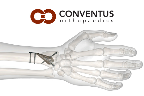
MINNEAPOLIS, Dec. 19, 2016 /PRNewswire/ — Conventus Orthopaedics, Inc. announced that over 700 patients now have been treated with the industry’s first expandable fracture cage, an innovative solution to fracture repairs in the upper extremity using a 3-dimensional approach. The Conventus Cage™, made of nitinol, a memory shape alloy, expands within the bone creating robust fixation and preventing further collapse of the fracture by providing a rigid internal scaffold upon completion of the procedure.

Conventus currently has five FDA-cleared indications in the upper extremity and the company is pursuing additional indications in the lower extremity and other areas of the skeletal anatomy.  Clinical follow-up of the first cage implantations is over four years now, showing not only excellent fracture repair but also fewer complications and reduced pain with faster recoveries for the patients.

“For years, surgeons have expressed the need for more versatile and reliable surgical options to repair simple to complex fractures,” said Paul Buckman, Conventus Orthopaedics CEO. “The Conventus Cages expand what is possible in patient care by enabling surgeons to repair a wider variety of fractures through less invasive means.”  Eric Fulkerson, M.D. of Muir Orthopaedics, Walnut Creek, CA said, “The Conventus Cage offers a solution that delivers reliably stable fracture repairs with consistently decreased recovery times and decreased pain for my patients. The innovative Cage design allows an expansion of fracture patterns treated with fixation as opposed to primary arthroplasty.” Since the Cage is implanted into bone, instead of onto bone, the potential for interference of hardware, as well as the need for a future surgery to remove fixation, is significantly reduced.  Andrew Schmidt, M.D., Chief of Orthopaedics, Hennepin County Medical Center, added, “For especially problematic fractures, such as those in the proximal humerus, the results after Cage fixation seem to be better, with more rapid return of motion and a dramatically lower incidence of surgical complications.”  Paul Paterson, M.D., orthopaedic surgeon in Buffalo, NY added, “The Conventus Cage offers tremendous stability of the fracture—better than plates and screws—through the use of 3-dimensional fixation. These fractures just do not move post repair.”

Conventus’ 3-dimensional Cage platform technology, emphasizes preservation of tissues and blood supply at the fracture site, creating a strong foundation by stabilizing bone fragments and facilitating early motion through less invasive procedures.

Conventus continues to expand their product portfolio and surgical applications in their quest to provide solutions throughout the entire musculoskeletal system.

For more information regarding use of the device please refer to www.conventusortho.com.


About Conventus Orthopaedics, Inc.
Conventus Orthopaedics, Inc. was founded by a team of medical device professionals and physicians with the sole purpose of improving patient care by creating a platform technology with less invasive solutions to a broad range of challenging periarticular fractures. Taking its name from the Latin word for coming together or union, Conventus Orthopaedics is focused on creating less invasive solutions for fractures in and around the joints. The company is dedicated to working with surgeons to improve healing and enable their patients’ return to normal activities.


SOURCE Conventus Orthopaedics, Inc.