Special Protein in Zebrafish May Lead to Full Spinal Cord Repair

Nov 04, 2016 – By Rhenn Anthony Taguiam


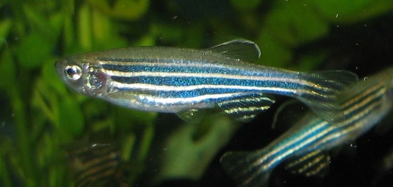
Researchers have discovered that zebrafish — the two-buck wonders in pet stores — may actually hold the key to do full spinal cord repair. It seems the zebrafish is capable of healing a completely severed spinal cord with special proteins.

An injury of this kind in humans can be paralyzing and can even lead to fatal conditions.

Researchers from Duke University have discovered that zebrafish possess a special protein that allows them to heal their severed spine. According to Medical Xpress, the study, published in Science, could lead to developments in tissue repair in humans.

Duke University’s Kenneth Poss claimed this is one of “nature’s most remarkable feats of regeneration.” This is because its potential implications and usage in the world of medicine is astounding. There are currently very limited methods of tissue repair, and animals like zebrafish may hold the key.

A zebrafish’s severed spinal cord forms a literal “bridge” that connects the gap in the injury. Cells form and extend to distances 10 times their own length to heal themselves. By eight weeks, the animals may have already fully reversed their paralysis.

Poss and his team conducted “molecular fishing” to find out which genes are responsible for such activity. It appears the CTGF or the connective tissue growth factor increases while the supporting cells appear to repair the injury.


READ THE REST HERE


InVivo Therapeutics Announces Appointment of Jeffrey Hatfield to Board of Directors

November 02, 2016

CAMBRIDGE, Mass.–(BUSINESS WIRE)–InVivo Therapeutics Holdings Corp. (NVIV) today announced the appointment of Jeffrey Hatfield to its Board of Directors. Mr. Hatfield most recently served as President, Chief Executive Officer, and Board Member for Vitae Pharmaceuticals and served in those capacities since joining the company in March 2004. Mr. Hatfield funded the company privately from 2005 to 2014 via three separate partnering transactions and took Vitae Pharmaceuticals public in 2014. On September 14, 2016, Allergan Plc announced that it had entered into a definitive agreement to purchase Vitae Pharmaceuticals for approximately $639 million in cash.

Prior to joining Vitae Pharmaceuticals, Mr. Hatfield worked at Bristol-Myers Squibb (BMS) in a variety of executive positions, including: Senior Vice President of BMS’ Immunology and Virology Divisions, where he was responsible for all aspects of the $1B annual revenue division; President and General Manager, BMS-Canada; and Vice President, U.S. Managed Health Care. Mr. Hatfield holds an M.B.A. from The Wharton School of the University of Pennsylvania and a bachelor’s degree in pharmacy from Purdue University, where he is a Distinguished Alumnus and adjunct professor. He has served on the Board of Ambit Biosciences (AMBI) before its acquisition by Daiichi-Sankyo, and is currently a member of the Board of Directors of the Biotechnology Industry Organization (BIO), serving on the Executive Committee of the Emerging Company Section. He is also a member of the advisory committees for Purdue University’s College of Pharmacy, Drexel University’s LeBow College of Business, and the Chapman-KGI School of BioPharmacy.

Mark Perrin, InVivo’s CEO and Chairman, said, “The Board of Directors is pleased to welcome Jeff. His invaluable experience in steering a company to clinical trials, managing successful product launches, business development transactions, and driving valuation leading to corporate acquisition makes him a great asset to the Board.”

“I’m personally very motivated and excited by the company’s work, as I’ve had direct experience with a family member who suffered a serious spinal cord injury at an early age,” Hatfield said. “I’m hopeful that Mark and the InVivo team can make a meaningful difference to the lives of those suffering these kinds of traumatic injury.”

About InVivo Therapeutics

InVivo Therapeutics Holdings Corp. is a research and clinical-stage biomaterials and biotechnology company with a focus on treatment of spinal cord injuries. The company was founded in 2005 with proprietary technology co-invented by Robert Langer, Sc.D., Professor at Massachusetts Institute of Technology, and Joseph P. Vacanti, M.D., who then was at Boston Children’s Hospital and who now is affiliated with Massachusetts General Hospital. In 2011, the company earned the David S. Apple Award from the American Spinal Injury Association for its outstanding contribution to spinal cord injury medicine. In 2015, the company’s investigational Neuro-Spinal Scaffoldreceived the 2015 Becker’s Healthcare Spine Device Award. The publicly-traded company is headquartered in Cambridge, MA. For more details, visit www.invivotherapeutics.com.

Contacts

InVivo Therapeutics Holdings Corp.
Brian Luque, 617-863-5535
Investor Relations
bluque@invivotherapeutics.com

Orthocell Receives Approval for Human Nerve Regeneration Study Using CelGro®

October 17, 2016

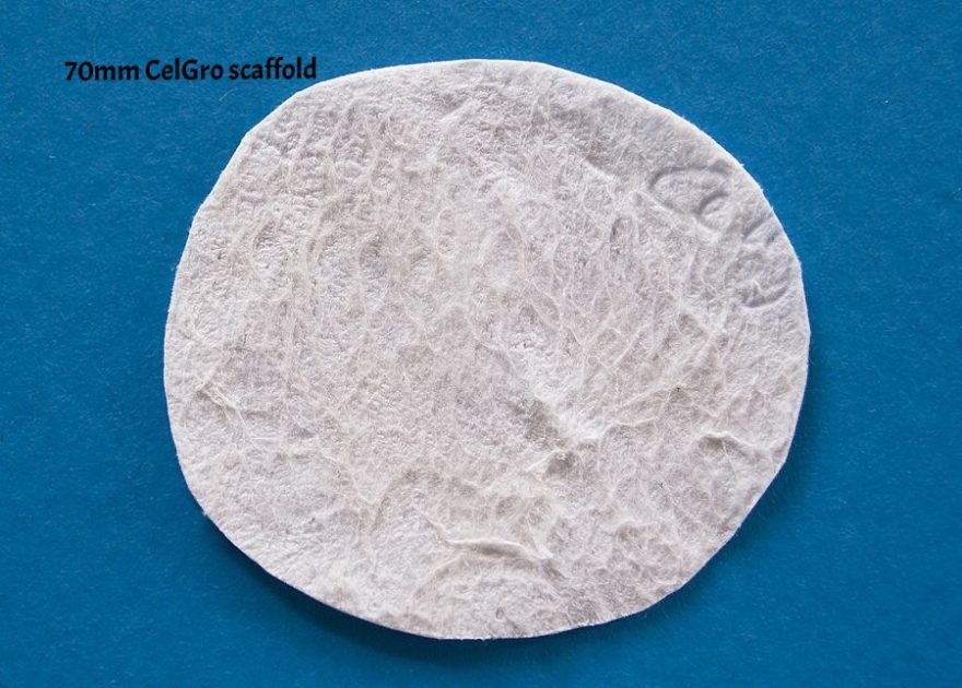
PERTH, Australia–(BUSINESS WIRE)–Regenerative medicine company Orthocell has been granted ethics approval for a human study examining the effectiveness of its CelGro® collagen medical device, to guide and promote nerve regeneration in severed peripheral nerves of the hand and upper limb.

The primary objective of this study is to demonstrate the safety, tolerability and effectiveness of CelGro® when used as a conduit in the surgical repair of peripheral nerve injuries. The study will involve 20 patients who have suffered injury to one or more peripheral nerves and will be undertaken at St John of God Hospital beginning in the fourth quarter of calendar 2016.

Peripheral nerve injury is most commonly caused by accidents or other trauma and in the US alone, over 20 million people are affected each year, at a cost of approximately $150 billion in annual health care dollars.

Principal investigator, orthopaedic surgeon Dr Alex O’Beirne said: “This is an exciting development that has the potential to improve patient outcomes by allowing for a suture-less repair to the damaged nerve, to guide nerve regeneration and accelerate the healing process.”

CelGro® is a biological medical device used as a scaffold for a variety of orthopaedic and general reconstructive surgical applications. Orthocell is also undertaking clinical studies using CelGro® to augment repair of the rotator cuff tendon within the shoulder, as an augment to guide and promote bone regeneration, as well as an augment to cartilage repair within the hip joint.

Orthocell Managing Director Paul Anderson said, “This is an exciting new phase in the development of CelGro® and its use as an augment to improve nerve repair. It further validates CelGro as a unique platform technology for soft tissue repair.”

Orthocell has submitted CelGro® for first regulatory approval in Europe and expects to receive notice of approval of its CE Mark application in 2016. Receipt of this approval will enable sales of CelGro® to commence in Europe, and trigger applications for other regulatory approvals in the United States, Australia and Japan in 2017.

About Orthocell Limited

Orthocell is a commercial-stage, regenerative medicine company focused on regenerating mobility for patients and our ageing population by developing products for a variety of tendon, cartilage and soft tissue injuries. Orthocell’s portfolio of products include TGA-approved stem cell therapies Autologous Tenocyte Implantation (Ortho-ATI™) and Autologous Chondrocyte Implantation (Ortho-ACI™), which aim to regenerate damaged tendon and cartilage tissue. The Company’s other major product is CelGro™, a collagen medical device which facilitates tissue repair and healing in a variety of orthopaedic, reconstructive and surgical applications and is being readied for first regulatory approvals.

Contacts

Orthocell Limited
General enquiries
Paul Anderson, +61 8 9360 2888
Managing Director
paulanderson@orthocell.com.au
or
Investor and Media enquiries
WE Buchan
Ben Walsh, +61 411 520 012
bwalsh@buchanwe.com.au

InVivo Therapeutics Announces Foothills Medical Centre in Calgary, Alberta as New Canadian Site for INSPIRE Study

October 19, 2016

CAMBRIDGE, Mass.–(BUSINESS WIRE)–InVivo Therapeutics Holdings Corp. (NVIV) today announced that the Foothills Medical Centre in Calgary, Alberta has been added as a Canadian clinical site for The INSPIRE Study: InVivo Study of Probable Benefit of theNeuro-Spinal Scaffold™ for Safety and Neurologic Recovery in Subjects with Complete Thoracic AIS A Spinal Cord Injury. The Foothills Medical Centre is the largest hospital in Alberta and is one of the most recognized medical facilities in Canada, providing advanced healthcare services to over two million people from Calgary and southern Alberta, the northwestern United States, southeastern British Columbia, and southern Saskatchewan.

“InVivo’s Neuro-Spinal Scaffold is one of the more innovative approaches to treating acute spinal cord injury in recent history and we look forward to being a part of the INSPIRE study,” said Steven Casha, M.D., Ph.D., Assistant Professor of Neurosurgery in the Department of Clinical Neurosciences and Principal Investigator at the study site.

Mark Perrin, InVivo’s CEO and Chairman, said, “We are pleased to welcome Dr. Casha and his team at the Foothills Medical Centre to the INSPIRE study. We will continue to expand our footprint in both the US and Canada.”

A new CEO’s Perspective discussing the current status of the INSPIRE study can be found on the InVivo Therapeutics website:http://www.invivotherapeutics.com/about-invivo/ceo-perspective/

There are now 24 clinical sites participating in the clinical study:

  • Banner University Medical Center, Tucson, AZ
  • Barnes-Jewish Hospital at Washington University Medical Center, St. Louis, MO
  • Ben Taub Hospital/Baylor College of Medicine, Houston, TX
  • Barrow Neurological Institute – St. Joseph’s Hospital and Medical Center, Phoenix, AZ
  • Carolina Neurosurgery and Spine Associates/Carolinas Rehabilitation, Charlotte, NC
  • Cooper Neurological Institute, Camden, NJ
  • Foothills Medical Centre, Calgary, Alberta, Canada
  • Goodman Campbell Brain and Spine/Indiana University Health Neuroscience Center, Indianapolis, IN
  • Hospital of the University of Pennsylvania, Philadelphia, PA
  • Keck Hospital of University of Southern California, Los Angeles, CA
  • Medical College of Wisconsin/Froedtert Hospital, Milwaukee, WI
  • Mount Sinai Hospital, New York, NY
  • Northwestern Medicine, Chicago, IL
  • Oregon Health & Science University, Portland, OR
  • Rutgers New Jersey Medical School, Newark, NJ
  • Thomas Jefferson University Hospital, Philadelphia, PA
  • Toronto Western Hospital, Toronto, ON, Canada
  • University of California, Davis Medical Center, Sacramento, CA
  • University of California, San Diego Medical Center, San Diego, CA
  • University of Kansas Medical Center, Kansas City, KS
  • University of Louisville Hospital, Louisville, KY
  • University of Pittsburgh Medical Center Presbyterian, Pittsburgh, PA
  • University of Virginia Health System, Charlottesville, VA
  • Vidant Medical Center, Greenville, NC

For more information, please visit the company’s ClinicalTrials.gov registration site: http://clinicaltrials.gov/ct2/show/study/NCT02138110

About the Neuro-Spinal Scaffold™ Implant

Following acute spinal cord injury, surgical implantation of the biodegradable Neuro-Spinal Scaffold within the decompressed and debrided injury epicenter is intended to support appositional healing, thereby reducing post-traumatic cavity formation, sparing white matter, and allowing neural regeneration across the healed wound epicenter. The Neuro-Spinal Scaffold, an investigational device, has received a Humanitarian Use Device (HUD) designation and currently is being evaluated in the INSPIRE pivotal probable benefit study for the treatment of patients with complete (AIS A) traumatic acute spinal cord injury.

About InVivo Therapeutics

InVivo Therapeutics Holdings Corp. is a research and clinical-stage biomaterials and biotechnology company with a focus on treatment of spinal cord injuries. The company was founded in 2005 with proprietary technology co-invented by Robert Langer, Sc.D., Professor at Massachusetts Institute of Technology, and Joseph P. Vacanti, M.D., who then was at Boston Children’s Hospital and who now is affiliated with Massachusetts General Hospital. In 2011, the company earned the David S. Apple Award from the American Spinal Injury Association for its outstanding contribution to spinal cord injury medicine. In 2015, the company’s investigational Neuro-Spinal Scaffoldreceived the 2015 Becker’s Healthcare Spine Device Award. The publicly-traded company is headquartered in Cambridge, MA. For more details, visit www.invivotherapeutics.com.

Safe Harbor Statement

Any statements contained in this press release or the CEO Perspective referred to herein that do not describe historical facts may constitute forward-looking statements within the meaning of the federal securities laws. These statements can be identified by words such as “believe,” “anticipate,” “intend,” “estimate,” “will,” “may,” “should,” “expect,” “designed to,” “potentially,” and similar expressions, and include statements regarding the safety and effectiveness of the Neuro-Spinal Scaffold, the expected timing of full enrollment in the INSPIRE study, the timing of the submission of the Humanitarian Device Exemption (HDE), the timing for opening of the pilot cervical SCI study, and the company’s anticipated cash balance. Any forward-looking statements contained herein are based on current expectations, and are subject to a number of risks and uncertainties. Factors that could cause actual future results to differ materially from current expectations include, but are not limited to, risks and uncertainties relating to the company’s ability to successfully open additional clinical sites for enrollment and to enroll additional patients; the ability to complete the INSPIRE study and submit an HDE; the company’s ability to receive regulatory approval for the Neuro-Spinal Scaffold; the company’s ability to commercialize its products; the company’s ability to develop, market and sell products based on its technology; the expected benefits and efficacy of the company’s products and technology in connection with the treatment of spinal cord injuries; the availability of substantial additional funding for the company to continue its operations and to conduct research and development, clinical studies and future product commercialization; and other risks associated with the company’s business, research, product development, regulatory approval, marketing and distribution plans and strategies identified and described in more detail in the company’s Annual Report on Form 10-K for the year ended December 31, 2015, and its other filings with the SEC, including the company’s Form 10-Qs and current reports on Form 8-K. The company does not undertake to update these forward-looking statements.

Contacts

InVivo Therapeutics
Brian Luque, 617-863-5535
Investor Relations
bluque@invivotherapeutics.com

Mainstay Medical ReActiv8-A sustains performance at one year in clinical trial

27th September 2016

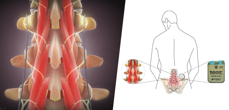
Mainstay Medical has announced the one-year results from the ReActiv8-A Clinical Trial, an international, multi-centre, prospective, single arm trial for ReActiv8 in people with disabling chronic low back pain and few other treatment options.

The one-year results show sustained performance in the ReActiv8-A Clinical Trial at the one-year follow-up with a clinically important, statistically significant and lasting improvement in the study’s key endpoints for pain (NRS), disability (ODI) and quality of life (EQ-5D).

Peter Crosby, chief executive officer of Mainstay, says, “We are very encouraged to see such strong and lasting benefits in this difficult-to-treat population. After one year of ReActiv8 treatment, 88% of subjects reported a clinically important improvement in one or more of the study endpoints, 81% were satisfied or very satisfied with the treatment and the majority continued to use the ReActiv8 treatment.”

The results presented are based on data from the first 47 subjects implanted in the ReActiv8-A Trial of whom 46 have completed the 90-day follow-up, 45 the 180-day follow-up and 41 the one-year follow-up.

To facilitate future comparison of results in the ReActiv8-A and the ReActiv8-B trial, all outcomes are presented relative to the data collected at the enrolment visit, according to a company release.

Results for all subjects at 90 days, 180 days and 1 year respectively are:

  • Ninety-three per cent, 87% and 88% with clinically important improvement in one or more of the study’s key endpoints.
  • Sixty-three per cent, 58% and 56% with clinically important improvement in low back pain NRS on the day.
  • Fifty per cent, 53% and 59% with clinically important improvement in ODI.
  • Eighty-nine per cent, 82% and 80% with clinically important improvement in EQ-5D.
  • Sixty-one per cent, 67% and 62% reported>50% pain relief.
  • Eighty-nine per cent, 84% and 81% were satisfied with ReActiv8 treatment.

The results for EQ-5D and ODI previously announced were relative to data collected at the pre-implant visit to 90 days and 180 days and were:

  • Fifty-sevent per cent and 60% with clinically important improvement in ODI.
  • Sixty-seven per cent and 73% with clinically important improvement in EQ-5D.

Adverse events incidence and type were comparable to those in clinical trials reported for other neurostimulation devices, according to the release, with no unanticipated adverse events, and no serious adverse events related to the device, therapy or procedure.

Trial offers hope of a treatment for spinal muscular atrophy

September 20, 2016 – University of Oxford

SMA occurs when people lack a gene called survival motor neuron 1 (SMN1). It can affect children in the womb or adults. This makes them unable to produce enough SMN protein, resulting is motor neuron degeneration and increasing muscle weakness. However, people have an almost identical gene called SMN2.

Existing proposed treatments are based on altering SMN2 to include a crucial part that is found in SMN1, enabling the production of SMN protein. This uses a splice-switching oligonucleotide or SSO. However, the difficulties of getting SSOs across the blood-brain barrier and into the central nervous system mean that they have to be injected into the spine with a lumbar puncture.

Researcher Dr Suzan Hammond explained: ‘Intrathecal delivery — injection around the spinal cord — makes a treatment less straightforward. Around a third of patients experience side effects. An additional complication is that SMA frequently leads to scoliosis — twisting of the spine — which can restrict such injections.’

The team at Oxford’s Department of Physiology, Anatomy and Genetics developed a treatment called Pip6a-PMO, in which the SSO was delivered using a peptide called Pip6a.

Dr Hammond explained: ‘Pip6a is highly effective at delivering SSOs to a wide variety of tissue in the body. We have confirmed that it can also get them into the brain and spinal cords in young and adult mice.’

When young mice — known as pups — with genetically engineered SMA were injected with the Pip6a-PMO, the results were rapidly clear: At just seven days old they were noticeably heavier and faster growing than untreated pups; at 12 days, tests found the treated pups much stronger than untreated counterparts. They also lived much longer, a median 167 days for mice treated with one dose of 10 microgrammes per gram of weight of Pip6a-PMO, compared to untreated pups’ 12 days.

Tests also found that two such doses of Pip6a-PMO markedly improved survival — with all mice treated in this way surviving at least 200 days and median survival of 457 days, 38 times longer than untreated mice and nearly three times longer than those who received a single dose.

Study of neuromuscular junctions, where motor neurons connect to muscles, showed that the effect of SMA, which destroys nerves at the junctions, was reversed by a single dose of the treatment, returning the connections to normal levels.

Professor Matthew Wood said: ‘While Pip6a was initially designed for Duchenne muscular dystrophy, we have shown that it can also be highly effective in SMA treatment. The survival of mice in this trial was far longer than any other treatment. The advantage is that it is both a central nervous system treatment and a systemic treatment for the wider body. Such an approach could also work for diseases like Parkinson’s, Huntingdon’s and ALS, and our focus will be extending the clinical applications of Pip-PMOs.’

The team are currently planning a 2-year study that would start next year, to evaluate this treatment in patients.


Story Source:

The above post is reprinted from materials provided byUniversity of Oxford. Note: Content may be edited for style and length.


Journal Reference:

  1. Suzan M. Hammond, Gareth Hazell, Fazel Shabanpoor, Amer F. Saleh, Melissa Bowerman, James N. Sleigh, Katharina E. Meijboom, Haiyan Zhou, Francesco Muntoni, Kevin Talbot, Michael J. Gait, Matthew J. A. Wood. Systemic peptide-mediated oligonucleotide therapy improves long-term survival in spinal muscular atrophy.Proceedings of the National Academy of Sciences, 2016; 201605731 DOI: 10.1073/pnas.1605731113

Cite This Page:

University of Oxford. “Trial offers hope of a treatment for spinal muscular atrophy.” ScienceDaily. ScienceDaily, 20 September 2016. <www.sciencedaily.com/releases/2016/09/160920154551.htm>.

Graphene nanoribbons show promise for healing spinal injuries

September 20, 2016 – Rice University

The Tour lab has spent a decade working with graphene nanoribbons, starting with the discovery of a chemical process to “unzip” them from multiwalled carbon nanotubes, as revealed in a Nature paper in 2009. Since then, the researchers have used them to enhance materials for the likes of deicers for airplane wings, better batteries and less-permeable containers for natural gas storage.

Now their work to develop nanoribbons for medical applications has resulted in a material dubbed Texas-PEG that may help knit damaged or even severed spinal cords.

A paper on the results of preliminary animal-model tests appears in the journal Surgical Neurology International.

Graphene nanoribbons customized for medical use by William Sikkema, a Rice graduate student and co-lead author of the paper, are highly soluble in polyethylene glycol (PEG), a biocompatible polymer gel used in surgeries, pharmaceutical products and in other biological applications. When the biocompatible nanoribbons have their edges functionalized with PEG chains and are then further mixed with PEG, they form an electrically active network that helps the severed ends of a spinal cord reconnect.

“Neurons grow nicely on graphene because it’s a conductive surface and it stimulates neuronal growth,” Tour said.

In experiments at Rice and elsewhere, neurons have been observed growing along graphene.

“We’re not the only lab that has demonstrated neurons growing on graphene in a petri dish,” he said. “The difference is other labs are commonly experimenting with water-soluble graphene oxide, which is far less conductive than graphene, or nonribbonized structures of graphene.

“We’ve developed a way to add water-solubilizing polymer chains to the edges of our nanoribbons that preserves their conductivity while rendering them soluble, and we’re just now starting to see the potential for this in biomedical applications,” he said. He added that ribbonized graphene structures allow for much smaller amounts to be used while preserving a conductive pathway that bridges the damaged spinal cords. Tour said only 1 percent of Texas-PEG consists of nanoribbons, but that’s enough to form a conductive scaffold through which the spinal cord can reconnect.

Texas-PEG succeeded in restoring function in a rodent with a severed spinal cord in a procedure performed at Konkuk University in South Korea by co-authors Bae Hwan Lee and C-Yoon Kim. Tour said the material reliably allowed motor and sensory neuronal signals to cross the gap 24 hours after complete transection of the spinal cord and almost perfect motor control recovery after two weeks.

“This is a major advance over previous work with PEG alone, which gave no recovery of sensory neuronal signals over the same period of time and only 10 percent motor control over four weeks,” Tour said.

The project began when Sikkema read about work by Italian neurosurgeon Sergio Canavero. Sikkema thought nanoribbons might enhance research that depended on PEG’s ability to promote the fusion of cell membranes by adding electrical conductivity and directional control for neurons as they spanned the gap between sections of the spinal cord. Contact with the doctor led to a collaboration with the South Korean researchers.

Tour said Texas-PEG’s potential to help patients with spinal cord injuries is too promising to be minimized. “Our goal is to develop this as a way to address spinal cord injury. We think we’re on the right path,” he said.

“This is an exciting neurophysiological analysis following complete severance of a spinal cord,” Tour said. “It is not a behavioral or locomotive study of the subsequent repair. The tangential singular locomotive analysis here is an intriguing marker, but it is not in a statistically significant set of animals. The next phases of the study will highlight the locomotive and behavioral skills with statistical relevance to assess whether these qualities follow the favorable neurophysiology that we recorded here.”


Story Source:

The above post is reprinted from materials provided by Rice University. Note: Content may be edited for style and length.


Journal Reference:

  1. JamesM Tour, BaeHwan Lee, C-Yoon Kim, WilliamK. A. Sikkema, In-Kyu Hwang, Hanseul Oh, UnJeng Kim. Spinal cord fusion with PEG-GNRs (TexasPEG): Neurophysiological recovery in 24 hours in rats. Surgical Neurology International, 2016; 7 (25): 632 DOI:10.4103/2152-7806.190475

Don’t Forget the Pulses! Aortoiliac Peripheral Artery Disease Masquerading as Lumbar Radiculopathy—A Report of 3 Cases

Am J Orthop. 2016 July

Lumbar radiculopathy is a common problem encountered by orthopedic surgeons, and typically presents with lower back or buttock pain radiating down the leg.1 While the most common causes of lumbar radiculopathy are lumbar disc herniation and spinal stenosis, the differential diagnosis for lower extremity pain is broad and can be musculoskeletal, vascular, neurologic, or inflammatory in nature.1,2Differentiating between orthopedic, neurologic, and vascular causes of leg pain, such as peripheral artery disease (PAD), can sometimes be challenging. This is especially true in aortoiliac PAD, which can present with hip, buttock, and thigh pain. Dorsalis pedis pulses can be palpable due to collateral circulation. A careful history and physical examination is crucial to the correct diagnosis. The history should clearly document the nature of the pain, details of walking impairment, and the alleviating effects of standing still or positional changes. A complete neurovascular examination should include observations regarding the skin, hair, and nails, examination of dorsal pedis, popliteal, and femoral pulses in comparison to the contralateral side, and documentation of dural tension signs. Misdiagnoses can send the patient down a path of unnecessary tests, unindicated procedures, and ultimately, a delay in definitive diagnosis and treatment.1

To our knowledge, this is the first report on a series of patients with thigh pain initially diagnosed as radiculopathy who underwent unproductive diagnostic tests and procedures, and ultimately were given delayed diagnoses of aortoiliac PAD. The patients provided written informed consent for print and electronic publication of these case reports.

Case 1

An 81-year-old woman with a medical history notable for hypertension, hyperlipidemia, and stroke initially presented to an outside orthopedic institution with complaints of several months of lower back and right hip, thigh, and leg pain when walking. She did not report any history of night pain, weakness, or numbness. Examination at the time was notable for painful back extension, 4/5 hip flexion strength on the right compared to 5/5 on the left, but symmetric reflexes and negative dural tension signs. X-rays showed multilevel degenerative disc disease of the lumbar spine, and magnetic resonance imaging (MRI) showed a small L3/4 disc protrusion causing impingement of the L4 nerve root.

A transforaminal epidural steroid injection at the L4 level was performed with minimal resolution of symptoms. Several months later, right-sided intra-articular facet injections were performed at the L4/5 and L5/S1 levels, again with minimal relief of symptoms. At this point, the patient was sent for further physical therapy.

Over a year after symptom onset, the patient presented to our institution and was evaluated by a vascular surgeon. Physical examination was notable for 1+ femoral artery and dorsal pedis pulses on the right side, compared to 2+ on the left. An aortoiliac duplex ultrasound showed severe significant stenosis of the right common iliac artery (>75%).


READ THE REST HERE

Improving the Lives of Patients, Bioness’ StimRouter™ Neuromodulation System Currently Being Implanted to Manage Chronic Pain

VALENCIA, Calif., Sept. 7, 2016 /PRNewswire/ — Bioness, Inc., the leading provider of cutting edge, clinically supported rehabilitation therapies, today announced a series of successful StimRouter Neuromodulation System implantations to manage chronic pain conditions originating from varied peripheral neuralgias. As a minimally invasive device designed to reduce pain by specifically targeting the affected peripheral nerve, StimRouter is designed to be a cost-effective alternative to injections, ongoing medication regiments, and complex surgeries.

Nearly 100 million Americans suffer from debilitating chronic pain, which may be experienced across all areas of the body including the arm, torso and leg. Chronic pain is often secondary to a primary condition (e.g. stroke) and can also result from entrapment or compression syndromes, post-surgical complications or failed surgeries.

“For many years we have had limited solutions to help our patients manage their debilitating pain,” shared William Porter McRoberts, MD, a trained Physiatrist and Interventional Spine and Pain Management Specialist based in Fort Lauderdale, Florida. “As hard as it is to understand the impact of one’s pain, it is easy to see when relief is delivered. I’m very pleased with how well my patients are responding to the StimRouter and believe that the technology will be suitable for a greater range of patients in the future.”

StimRouter was the first FDA cleared non-drug, long-term, minimally invasive neuromodulation medical device indicated to treat chronic pain of a peripheral nerve origin. The patient controlled device is an adjunct to other modes of therapy (e.g., medications) and is being well received by patients and clinicians alike.

The StimRouter is currently being implanted at prestigious clinical institutions across the country to treat chronic peripheral nerve pain, with specific focus on the following conditions or areas:

  • Axillary nerve (e.g. post-stroke shoulder pain)
  • Ulnar nerve (e.g. cubital tunnel syndrome)
  • Ilioinguinal (e.g. post-surgical hernia complication)
  • Superior Cluneal nerve (e.g. lower back neuralgia)

“As clinicians and patients continue to look for effective and sustainable ways to treat and manage pain, it is rewarding to see patients thrive with our StimRouter technology,” shared Todd Cushman, President and CEO of Bioness. “Pain is more than just a barrier to a comfortable life and pursing employment, it can be what prevents patients from being able to seek therapy to rehabilitate an injury or illness.”

For more information on the StimRouter as well as videos of real patients sharing their StimRouter experience, please visit www.stimrouter.com.

About StimRouter Neuromodulation System
StimRouter is cleared by the FDA to treat chronic pain of peripheral nerve origin. StimRouter is a minimally invasive neuromodulation medical device consisting of a thin, implanted lead with conductive electrode, external pulse transmitter (EPT), and hand-held wireless patient programmer. Electrical signals are transmitted transdermally from the EPT through the electrode, down the lead to the target nerve. StimRouter is programmed at the direction of the physician to meet patient requirements but is controlled by the patient to address the patients specific, changing pain management needs.

About Bioness Inc.
Bioness is the leading provider of innovative technologies helping people regain mobility and independence. Bioness solutions include external and implantable functional electrical stimulation (FES) systems, robotic systems and software-based therapy programs providing functional and therapeutic benefits for individuals affected by pain, central nervous system disorders and orthopedic injuries. Individual results vary. Consult with a qualified physician to determine if this product is right for you. Contraindications, adverse reactions and precautions are available online at www.bioness.com

Media Relations Contact Information
Next Step Communications
bioness@nextstepcomms.com
781.326.1741

StimRouter™ and Bioness® are trademarks of Bioness, Inc. | www.bioness.com | Rx Only | Additional information about StimRouter can be found at www.stimrouter.com.


To view the original version on PR Newswire, visit:http://www.prnewswire.com/news-releases/improving-the-lives-of-patients-bioness-stimrouter-neuromodulation-system-currently-being-implanted-to-manage-chronic-pain-300323378.html

Nevro touts 2-year data on Senza spinal cord stim

 By 


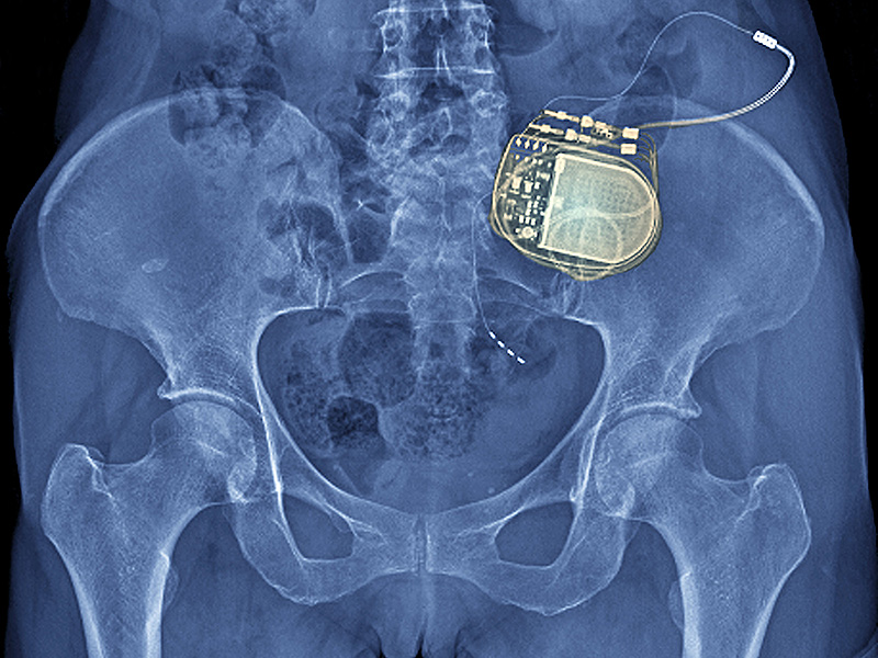
Nevro Corp. (NYSE:NVRO) today released 2-year results from Senza-RCT study of its HF10 spinal cord stimulation system, touting superior results over low-frequency SCS therapy treatments.

The Redwood City, Calif.-based company said results from the study were published in the journal Neurosurgery. Nevro said the trial is the largest prospective randomized trial of SCS systems and the 1st to evaluate the comparative effectiveness of spinal cord stimulation therapies.

The primary endpoint for the trial was a responder rate signifying a greater-than 50% reduction in back pain from the baseline at 3 months, with a secondary endpoint set at 12 months. Leg pain reductions and 24 month results from back pain reduction were also polled for secondary endpoints.

Patients treated with the company’s HF10 SCS reported significant improvements in superior back pain and leg pain 76.5% and 72.9% respectively, much higher than the 49.3% rates reported with traditional SCS for both back and leg pain.

Superior and durable pain relief results also indicated superiority for the HF10, with Visual Analog Scale scores for back and leg pain of 2.4 cm with HF10 versus 4.5 cm and 3.9 cm with traditional SCS, according to the study.


READ THE REST HERE