Rotation Medical Bioinductive Implant Induces New Tissue Formation in Patients with Partial-Thickness Rotator Cuff Tears

September 07, 2016

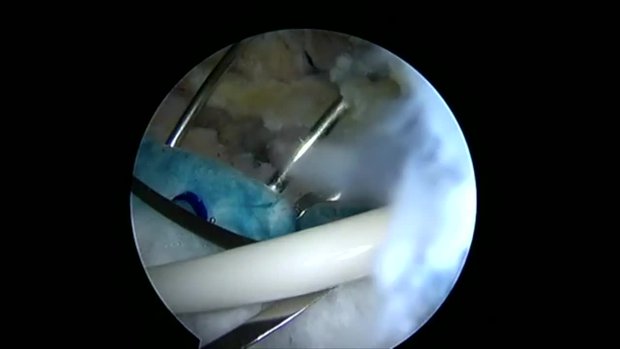
PLYMOUTH, Minn.–(BUSINESS WIRE)–Rotation Medical Inc., a medical device company focused on developing new technologies to treat rotator cuff disease, today announced that study results published in the current issue of Muscle, Ligaments and Tendons Journal showed that the company’s collagen-based bioinductive implant induced new tissue formation in all study patients with rotator cuff tears. The Rotation Medical rotator cuff system is a novel, implant-based solution for rotator cuff repair and a new alternative to traditional surgical repair.

The study assessed the ability of Rotation Medical’s bioinductive implant to induce new tissue formation and limit tear progression when placed on the bursal surface of partial thickness cuff tears. The implant induced significant new tissue formation in all 13 patients by three months (mean increase in tendon thickness 2.2 ± 0.26 mm), and the tissue matured over time and became radiologically indistinguishable from the underlying tendon. No tear progression was observed on MRI in any of the patients during the 24-month post-operative period. All patients’ Constant and American Shoulder and Elbow Society clinical scores improved significantly over time.1

“Partial-thickness rotator cuff tears frequently enlarge due to increased local strain and often progress to full-thickness tears,” said Dr. Desmond John Bokor, lead study investigator and associate professor in the Department of Orthopaedic Surgery at Macquarie University in Australia. “The results of this study demonstrate the ability of the bioinductive implant to induce new tendon-like tissue, enabling partial-thickness rotator cuff tears to decrease in size and in most cases disappear. The ability to heal partial-thickness rotator cuff defects, and thus prevent tear propagation and progressive tendon degeneration, represents a novel interventional treatment paradigm for these lesions.”

Rotator cuff damage is the most common source of shoulder pain, affecting more than 4 million people annually in the U.S. Traditional approaches to treating degenerate or torn rotator cuffs often do not address the poor quality of the underlying tendon tissue, and a significant number of these tendons, after standard treatment, either degenerate further and/or re-tear. Cleared by the U.S. Food and Drug Administration in March 2014, the Rotation Medical bioinductive implant is designed to address this limitation by inducing new tissue growth at the site of implantation, resulting in increased tendon thickness and healing of tendon defects with new tissue growth. The collagen-based implant is about the size of a postage stamp and it is part of the Rotation Medical rotator cuff system, which also includes disposable instruments that allow the arthroscopic procedure to be performed easily and quickly.

“This study is further evidence that the Rotation Medical rotator cuff system has the potential to transform the treatment of rotator cuff disease,” said Martha Shadan, president and CEO of Rotation Medical. “Our bioinductive implant addresses both the biomechanics and biology required to heal a rotator cuff tendon tear, preventing rotator cuff tears from becoming larger over time, reducing the incidence of re-tears and, in some cases, shortening patient recovery time.”

The study, “Evidence of healing of partial-thickness rotator cuff tears following arthroscopic augmentation with a collagen implant: a 2-year MRI follow-up,” adds to the growing body of literature supporting the use of the bioinductive implant as a novel treatment for rotator cuff partial thickness tears. Additional publications and information about the Rotation Medical rotator cuff system are available on the company’s website.

About the Study

The implant was inserted via arthroscopic surgery. A total of 13 patients with intermediate (3-6 mm) to high-grade (>6 mm) partial thickness cuff tears completed two years of follow-up. At three, six, 12, and 24 months postoperatively, tendon thickness, defect size and quality were evaluated using magnetic resonance imaging (MRI), and clinical outcomes were assessed using the Constant and American Shoulder and Elbow Society scores. The partial-thickness cuff tears showed consistent filling of the defects, with complete healing in seven patients at 12 months, and a progressive improvement in tendon quality in the remaining patients. No tear progression was observed by MRI in any of the patients at 24 months. All clinical scores improved significantly over time. At 24 months, 12 of 13 patients (92 percent) had satisfactory or better results.

About Rotation Medical

Rotation Medical Inc. was founded in 2009 and is committed to improving the treatment of rotator cuff disease with the Rotation Medical rotator cuff system, a breakthrough technology that has the potential to prevent rotator cuff disease progression and reduce re-tears by inducing the growth of new tendinous tissue. The company is privately held and funded by New Enterprise Associates (NEA), Life Sciences Partners (LSP) and Pappas Ventures. For more information, visit www.rotationmedical.com.

________________________
1 Bokor DJ, Sonnabend D, Deady L, Cass B, Young A, Van Kampen C, Arnoczky S. Evidence of healing of partial-thickness rotator cuff tears following arthroscopic augmentation with a collagen implant: a 2-year MRI follow-up. Muscle Ligaments Tendons J. 2016 May 19;6(1):16-25. doi: 10.11138/mltj/2016.6.1.016.

Contacts

Merryman Communications
Joni Ramirez, 323-532-0746
joni@merrymancommunications.com

3-D graphene has promise for bio applications, Scientists weld nanoscale sheets to form tough, porous material

September 2, 2016 – Ajayan Group/Rice University

The Rice lab of materials scientist Pulickel Ajayan and colleagues in Texas, Brazil and India used spark plasma sintering to weld flakes of graphene oxide into porous solids that compare favorably with the mechanical properties and biocompatibility of titanium, a standard bone-replacement material.

The discovery is the subject of a paper in Advanced Materials.

The researchers believe their technique will give them the ability to create highly complex shapes out of graphene in minutes using graphite molds, which they believe would be easier to process than specialty metals.

“We started thinking about this for bone implants because graphene is one of the most intriguing materials with many possibilities and it’s generally biocompatible,” said Rice postdoctoral research associate Chandra Sekhar Tiwary, co-lead author of the paper with Dibyendu Chakravarty of the International Advanced Research Center for Powder Metallurgy and New Materials in Hyderabad, India. “Four things are important: its mechanical properties, density, porosity and biocompatibility.”

Tiwary said spark plasma sintering is being used in industry to make complex parts, generally with ceramics. “The technique uses a high pulse current that welds the flakes together instantly. You only need high voltage, not high pressure or temperatures,” he said. The material they made is nearly 50 percent porous, with a density half that of graphite and a quarter of titanium metal. But it has enough compressive strength — 40 megapascals — to qualify it for bone implants, he said. The strength of the bonds between sheets keeps it from disintegrating in water.

The researchers controlled the density of the material by altering the voltage that delivers the highly localized blast of heat that makes the nanoscale welds. Though the experiments were carried out at room temperature, the researchers made graphene solids of various density by raising these sintering temperatures from 200 to 400 degrees Celsius. Samples made at local temperatures of 300 C proved best, Tiwary said. “The nice thing about two-dimensional materials is that they give you a lot of surface area to connect. With graphene, you just need to overcome a small activation barrier to make very strong welds,” he said.


READ THE REST HERE


MiMedx Announces Nationwide Launch of AmnioFill™

MARIETTA, Ga., Sept. 1, 2016 /PRNewswire/ — MiMedx Group, Inc. (NASDAQ: MDXG), the leading regenerative medicine company utilizing human amniotic tissue and patent-protected processes to develop and market advanced products and therapies for the Wound Care, Surgical, Orthopedic, Spine, Sports Medicine, Ophthalmic, and Dental sectors of healthcare, announced today its plans for the nationwide launch of AmnioFill™, the first product in the MiMedx placental collagen matrix product family to be commercially launched.

Physicians are in need of a product to treat larger acute and chronic wounds encountered in the surgical setting. Slated for nationwide release later this month, AmnioFill is being offered in multiple sizes and configurations to address this and other surgical needs.

AmnioFill is a collagenous matrix derived from the placenta and comprised of placental extracellular matrix (ECM) tissue. AmnioFill is a tissue allograft containing ECM proteins, growth factors, cytokines and other specialty proteins present in placental tissue. Over 226 growth factors, cytokines and chemokines, including important modulators of inflammation and factors critically important in wound healing, are contained in the AmnioFill placental tissue. MiMedx employs terminal sterilization in addition to aseptic processing techniques in its proprietary processing methodology to enhance the safety of AmnioFill and its other amniotic and placental products.

Parker H. “Pete” Petit, Chairman and CEO, said, “AmnioFill will be a great addition to our product lines, addressing the needs of both Wound Care and Surgical markets. For example, we expect that AmnioFill will be an ideal solution for physicians in the treatment of dehisced surgical wounds and other deep complex and hard-to-heal surgical wounds that require a connective tissue matrix to replace or supplement damaged or inadequate integumental tissue.”

Christopher M. Cashman, Executive Vice President and Chief Commercialization Officer, commented, “The historical costs to treat these types of wounds are significant, and the quality of life issues of non-resolved wounds of this nature can be devastating.  There were 53 million outpatient procedures performed in the United States in 2010. Despite advances in preoperative care, the rate of surgical wound dehiscence has not decreased in recent years with 1% to 3% of patients experiencing wound dehiscence. For example, breast reconstruction incisional dehiscence rates range from 10% to 15% in a setting with radiation therapy and abdominal wall surgical dehiscences have a mortality rate as high as 45%.”

Bill Taylor, President and COO, stated, “Our published scientific studies have demonstrated that our dehydrated Human Amnion/Chorion Membrane (dHACM) allografts cause stem cells to migrate and proliferate. Moreover, these scientific studies also demonstrated our dHACM allografts promote angiogenesis. Multiple clinical studies have confirmed that stem cell migration, proliferation and recruitment as well as angiogenesis are essential in wound healing.”

Cashman added, “AmnioFill is designed to provide a scaffold for recruited cells to attach, populate and proliferate. The placental tissues in the scaffold should modulate the activity of the recruited cells to generate new tissue for these larger acute and chronic surgical wounds. When used earlier in the treatment of these complex wounds, we believe AmnioFill becomes an even more cost effective approach as a step therapy for wound closure.”

About MiMedx

MiMedx® is an integrated developer, processor and marketer of patent protected and proprietary regenerative biomaterial products and bioimplants processed from human amniotic membrane and other birth tissues and human skin and bone.  “Innovations in Regenerative Biomaterials” is the framework behind our mission to give physicians products and tissues to help the body heal itself.  The MiMedx allograft product families include our: dHACM family with AmnioFix®, EpiFix® and EpiBurn® brands; Amniotic Fluid family with OrthoFlo brand; Umbilical family with EpiCord™ and AmnioCord™ brands; Placental Collagen family with CollaFix™ and AmnioFill™ brands; Bone family with Physio® brand; and Skin family with AlloBurn™ brand. AmnioFix,  EpiFix, and EpiBurn are our tissue technologies processed from human amniotic membrane; OrthoFlo is an amniotic fluid derived allograft;  EpiCord™ and AmnioCord™ are derived from the umbilical cord; Physio is a unique bone grafting material comprised of 100% bone tissue with no added carrier; AlloBurn is a skin product derived from human skin designed for the treatment of burns; and  CollaFix, our next brand we plan to commercialize, is our collagen fiber technology, developed with our patented cross-linking polymers, designed to mimic the natural composition, structure and mechanical properties of musculoskeletal tissues in order to augment their repair.

We process the human amniotic membrane utilizing our proprietary PURION® Process, to produce a safe and effective implant. MiMedx proprietary processing methodology employs aseptic processing techniques in addition to terminal sterilization.  MiMedx is the leading supplier of amniotic tissue, having supplied over 700,000 allografts to date for application in the Wound Care, Burn, Surgical, Orthopedic, Spine, Sports Medicine, Ophthalmic and Dental sectors of healthcare.

Safe Harbor Statement

This press release includes statements that look forward in time or that express management’s beliefs, expectations or hopes. Such statements are forward-looking statements within the meaning of the Private Securities Litigation Reform Act of 1995. These statements include, but are not limited to the Company’s belief that AmnioFill will be a great addition to its product lines, addressing the needs of both wound care and surgical markets; that AmnioFill will be an ideal solution for physicians in the treatment of dehisced surgical wounds and other deep complex and hard-to-heal surgical wounds that require a connective tissue matrix to replace or supplement damages or inadequate integumental tissue; and that, when used earlier in the treatment of complex wounds, AmnioFill becomes an even more cost effective approach as a step therapy for wound closure. Among the risks and uncertainties that could cause actual results to differ materially from those indicated by such forward-looking statements include that demand for, and acceptance of, any new product by the medical community may not be as expected; factors such as third party reimbursement may impact physician use of product; AmnioFill may not be used as anticipated or perform as anticipated in the clinical setting; AmnioFill may  not be as cost effective as anticipated, and the risk factors detailed from time to time in the Company’s periodic Securities and Exchange Commission filings, including, without limitation, its 10-K filing for the fiscal year ended December 31, 2015 and its most recent 10Q filing. By making these forward-looking statements, the Company does not undertake to update them in any manner except as may be required by the Company’s disclosure obligations in filings it makes with the Securities and Exchange Commission under the federal securities laws.

SOURCE MiMedx Group, Inc.

Related Links

http://www.mimedx.com