Tangible Solutions Aims to Achieve the Vision Behind “Industry 4.0” with Concept Laser

Grapevine, Texas, November 28, 2016  –Tangible Solutions has announced today that they will add 3D metal printing to their portfolio of capabilities. They will be integrating five Mlab cusing machines and one M2 cusing machine into their new 25,000 square feet facility in Fairborn, a suburb of Dayton, Ohio. This will render Tangible Solutions as one of the largest service providers of Concept Laser’s metal powder-bed technology in the country.

“We share Concept Laser’s vision for building a “smart factory” that supports the principles of Industry 4.0: automation, digitization and interoperability of various technologies within a factory. Concept Laser was the first and only 3D metal machine manufacturer to share a complete and tangible plan of helping organizations achieve industrial, serial production. We believe their technology roadmap will only make 3D metal printing more cost effective and flexible”, says Adam Clark and Chris Collins, Founders of Tangible Solutions.

“The team at Tangible Solutions are entrepreneurial and forward-looking; In only three years, they have made a positive impact in manufacturing in Ohio. We are committed to their success,” states John Murray, President and CEO of Concept Laser Inc.

Tangible Solutions was established in 2013 to offer services related to 3D printing, also known as additive manufacturing. Their offerings include 3D modeling and design, rapid prototyping, and developing course curriculum for education. The addition of metal 3D Printing in both reactive and non-reactive material allows them to broaden their reach with medical and aerospace customers. The Mlab cusing machines will be dedicated to printing with Titanium and the M2 cusing machine, nicknamed as the ‘workhorse’ for its stability and reliability, will print in variety of metal powder. The open architecture of Concept Laser’s machine portfolio allows customers the freedom to manufacture with any type of metal material; no restrictions are placed for using only Concept Laser’s metal powders.

Additive manufacturing involves taking digital designs from computer aided design (CAD) software, and laying horizontal cross-sections to manufacture the part. Additive components are typically lighter and more durable than traditional forged parts because they require less welding and machining. Because additive parts are essentially “grown” from the ground up, they generate far less scrap material. Freed of traditional manufacturing restrictions, additive manufacturing dramatically expands the design possibilities for engineers

Concept Laser recently made their vision of the “AM Factory of Tomorrow” a reality by releasing their new product called M LINE Factory which features a modular machine architecture with the ability to decouple pre-production from production. This allows factories to operate in parallel rather than sequentially, presenting economical series production.

ABOUT TANGIBLE SOLUTIONS

Tangible Solutions is a leading additive manufacturing company, providing prototype and contract manufacturing services, training and consulting, and research & development. Tangible Solutions services a wide variety of customers in the biomedical, aerospace, and automotive industries.

Tangible Solutions was established in 2013 to provide 3D printing and engineering design services. Quickly, Tangible became a leader in the Additive Manufacturing industry due to their knowledge about various 3D Printing methods and materials, quick turnaround times, and ability to produce professional products that held up to rigorous standards. Tangible Solutions also develops curriculum for workforce development that is in use today by government and academic institutions. The Tangible Solutions team is known for its subject matter expertise in Additive Manufacturing and dedication to customers.

ABOUT CONCEPT LASER

Concept Laser GmbH is one of the world’s leading providers of machine and plant technology for the 3D printing of metal components. Founded by Frank Herzog in 2000, the patented LaserCUSING® process – powder-bed-based laser melting of metals – opens up new freedom to configuring components and also permits the tool-free, economic fabrication of highly complex parts in fairly small batch sizes.

Concept Laser serves various industries, ranging from medical, dental, aerospace, toolmaking and mold construction, automotive and jewelry. Concept Laser machines are compatible with a diverse set of powder materials, such as stainless steel and hot-work steels, aluminum and titanium alloys, as well as precious metals for jewelry and dental applications.

Concept Laser Inc. is headquartered in Grapevine, Texas and is a US-based wholly owned subsidiary of Concept Laser GmbH. For more information, visit our website at www.conceptlaserinc.com

LaserCUSING® is a registered trademark of Concept Laser.

NuVasive to Showcase Integrated Global Alignment for the Cervical Spine at CSRS 2016

SAN DIEGO, CA–(Marketwired – December 01, 2016) – NuVasive, Inc. (NASDAQ: NUVA), a leading medical device company focused on transforming spine surgery with minimally disruptive, procedurally-integrated solutions, today announced the exhibiting of the Company’s recently launched Integrated Global Alignment™ (iGA) for the Cervical Spine at the Cervical Spine Research Society (CSRS) 44th Annual Meeting in Toronto. NuVasive recently expanded its iGA platform to support all spine procedures, including cervical alignment, making NuVasive the first company to offer a solution to address spinal alignment for all spine procedures.

“Our mission is to help surgeons achieve total sagittal alignment and deliver more predictable clinical outcomes for patients through global alignment of the spine,” said Jason Hannon, NuVasive’s president and chief operating officer. “With the newly expanded iGA platform, we expect to build off of the tremendous momentum we have seen with iGA to meet the needs of patients with varying cervical pathologies.”

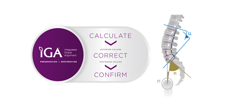
iGA is a proprietary, procedurally-integrated platform of specialized tools and software designed to help surgeons achieve more precise spinal column alignment. The expanded platform provides surgeons an intuitive software workflow to pre-plan their surgical procedure and then confirm achievement of the desired correction both intraoperatively and post-operatively. It also includes hardware tools, such as Bendini® OCT computer assisted rod bending technology, VuePoint® II OCT fixation system, and the CoRoent® Small Interlock Hyperlordotic system, that enhance the surgeon’s ability to achieve the intended spinal alignment. The newly launched CoRoent Small Interlock Hyperlordotic system allows surgeons to address cervical sagittal deformities from an anterior approach with interbodies that have up to 20 degrees of lordosis.

NuVasive is committed to a global approach for assessing, preserving and restoring spinal alignment in an effort to promote surgical efficiencies, lasting patient outcomes and improved quality of life. NuVasive’s differentiated technology helps surgeons:

  • Calculate the patient’s cervical alignment and preoperatively plan prior to surgery,
  • Correct and assess the cervical spine intraoperatively through integrated procedural solutions, and
  • Confirm postoperatively to reconcile the preoperative plan and the intraoperative assessment.

NuVasive CSRS Participation Details

NuVasive will showcase and demo its new technologies at NuVasive Booth #417 on the CSRS exhibit floor. In addition, the Company will host a CSRS Symposium where attendees will have the opportunity to understand the clinical rationale for restoring and/or preserving sagittal balance in the cervical spine. Following the presentation, faculty will demonstrate computer-assisted surgery techniques including case examples. Hands-on with the technologies will be available at the conclusion of the workshop.

DATE: Thursday, December 1 at 12:00 p.m. — 2:30 p.m.

LOCATION: Westin Harbour Castle — Yonge room

CSRS TOPIC: Cervical Spinal Deformity and Complex Osteotomies

TITLE: Cervical Alignment Matters™: Emerging Techniques to Restore Global Alignment with Computer-assisted Surgery

FACULTY: Donald J. Blaskiewicz, M.D., Themistocles Protopsaltis, M.D., and Neill M. Wright, M.D.

About NuVasive

NuVasive, Inc. (NASDAQ: NUVA) is a world leader in minimally invasive, procedurally-integrated spine solutions. From complex spinal deformity to degenerative spinal conditions, NuVasive is transforming spine surgery with innovative technologies designed to deliver reproducible and clinically proven surgical outcomes. NuVasive’s highly differentiated, procedurally-integrated solutions include access instruments, implantable hardware and software systems for surgical planning and reconciliation technology that centers on achieving the global alignment of the spine. With $811 million in revenues (2015), NuVasive has an approximate 2,200 person workforce in more than 40 countries around the world. For more information, please visit www.nuvasive.com.

Forward-Looking Statements

NuVasive cautions you that statements included in this news release that are not a description of historical facts are forward-looking statements that involve risks, uncertainties, assumptions and other factors which, if they do not materialize or prove correct, could cause NuVasive’s results to differ materially from historical results or those expressed or implied by such forward-looking statements. The potential risks and uncertainties which contribute to the uncertain nature of these statements include, among others, risks associated with acceptance of the Company’s surgical products and procedures by spine surgeons, development and acceptance of new products or product enhancements, clinical and statistical verification of the benefits achieved via the use of NuVasive’s products (including the iGA™ platform), the Company’s ability to effectually manage inventory as it continues to release new products, its ability to recruit and retain management and key personnel, and the other risks and uncertainties described in NuVasive’s news releases and periodic filings with the Securities and Exchange Commission. NuVasive’s public filings with the Securities and Exchange Commission are available at www.sec.gov. NuVasive assumes no obligation to update any forward-looking statement to reflect events or circumstances arising after the date on which it was made.

Contact Information

Investor Contact:
Suzanne Hatcher
NuVasive, Inc.
858-458-2240
Email contact

Media Contact:
Stefanie Mazer
NuVasive, Inc.
858-320-5243
Email contact

First AxioMed Viscoelastic Total Disc Replacement Implanted in Australia

BRISBANE, AUSTRALIA (PRWEB) NOVEMBER 29, 2016

AxioMed is pleased to announce the success of the first viscoelastic Freedom cervical case in Australia. Dr. Richard Laherty of Queensland Neurosurgery & Spine Surgery completed the procedure on Monday, November 28th at the Princess Alexandria Hospital in Brisbane, Australia. The procedure was performed on a 47-year-old male patient suffering from degenerative disc disease with radiculopathy, as a result of degenerative cervical discs at levels C5-7. The patient failed conservative treatments prior to undergoing surgery.

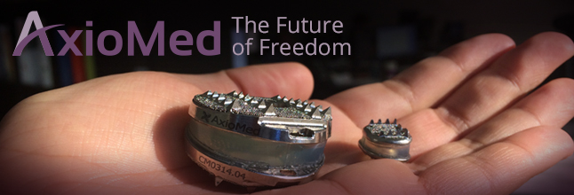
The AxioMed viscoelastic disc is a next-generation disc replacement that restores natural disc height, lordosis, stability, and motion in the human spine. AxioMed was approved in September to market and sell their viscoelastic cervical and lumbar Freedom total disc replacements in Australia by the Therapeutic Goods Administration.

Dr. Richard Laherty spoke to the advantages of the AxioMed cervical disc after the operation: “The Freedom cervical disc is extremely easy to implant and has a great anatomical fit for restoring disc height and lordosis in the cervical spines of my patients. I am excited to have this viscoelastic technology that mimics the natural motion of a healthy human disc, and to make this solution available to my patients. It will provide a faster surgical recovery time and increase a patient’s overall health and satisfaction.”

AxioMed CEO Dr. Kingsley Chin explained how an experience like Dr. Laherty’s aligns with AxioMed’s vision: “AxioMed believes it can replicate the success of total joint restoration in the spine with our innovative and advanced viscoelastic total disc replacements with a high degree of patient satisfaction.”

Dr. Chin added, “With the addition of the lateral lumbar technique, we expect AxioMed to be the worldwide leader in disc replacement surgery.”

Dr. Laherty currently practices general neurosurgery with a special interest in minimally invasive surgical techniques for management of complex spine conditions. Dr. Laherty graduated from University of Queensland and completed postgraduate fellowship training at Princess Alexandra Hospital, Brisbane and St. Vincents and Concorde Hospitals in Sydney. He is extensively involved in both research and training young neurosurgeons in the latest technologies via his roles at both Princess Alexandra Hospital and the University of Queensland.

About AxioMed

Founded in 2001, AxioMed (http://www.axiomed.com/) began its journey of exhaustively proving the Freedom® Disc through clinical studies in the USA and Europe, research, development and testing. In 2014, KICVentures recognized the disc’s enormous potential and acquired the company into their healthcare portfolio. AxioMed owns an exclusive viscoelastic material license on its proprietary Freedom Disc technology.

COA 2016 in Beijing, China: joimax® Shows Major Presence and Inaugurates New NASS-joimax® MISS Training Centre

Joimax®, the global acting German developer and marketer of technologies and training methods for endoscopic minimally invasive spinal surgery, again exhibited at Chinese OrthopedicAssociation (COA), which took place Nov. 17th to 20th, 2016 at China National Convention Center in Beijing, China. During the exhibition, the new NASS – joimax® MISS Training Centre was inaugurated.

The 18th Chinese Orthopedic Association Annual Meeting and the 11th International Congress of Chinese Orthopedic Association (COA) this year has again taken place in Beijing and was visited by more than 35,000 surgeons who gathered for the latest research results and up-to-date information on technology and clinical progression in orthopedics including spine surgery. joimax® had a major presence with a large booth showing its endoscopic minimally invasive product range. Furthermore on the two major meeting days, 30 lectures from well-known experts were given at its booth.

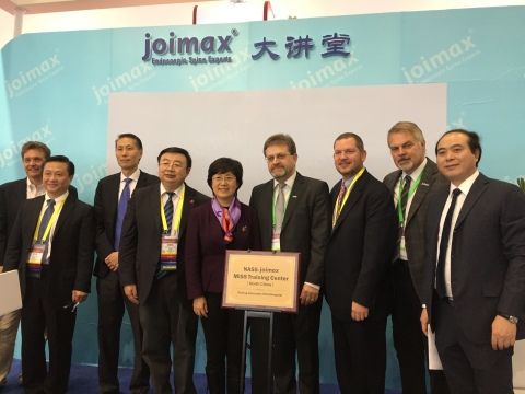
During the COA, the new NASS-joimax® MISS Training Centre in China was inaugurated on Friday Nov. 18th, 2016. The ceremony took place at the joimax® booth together with leaders from the Peking University Third Hospital and members of the NASS Board of Directors. The Chinese market is a very important one for joimax® because Chinese surgeons are highly interested in adopting and deepening their knowledge in endoscopic minimally invasive techniques. joimax® is playing a major role in advanced training and is a driving power in the field of continuing education.

“With the launch of TESSYS® ISeeUTM, which has been newly designed especially for the Chinese market, joimax®offers an even more simplified access under facilitated orientation,” says Wolfgang Ries, CEO and founder of joimax®. “The existing TESSYS® trays can furthermore be upgraded with the TESSYS® ISeeUTM supplements,” he continues.

Moreover, joimax® also presented its latest product, the newly launched Intracs® Interaoperative Navigation Tracking & Control System for the first time in China.

About joimax®

Founded in Karlsruhe, Germany, in 2001, joimax® is the leading developer and marketer of complete systems for endoscopic minimally invasive spinal surgery. With TESSYS® (transforaminal), iLESSYS® (interlaminar) and CESSYS®(cervical) for decompression procedures, MultiZYTE® RT (e.g. for rhizotomy) and with MultiZYTE® SI for SI-Joint therapy or with EndoLIF® and Percusys® for endoscopic minimally-invasive assisted stabilizations, proven endoscopic systems are provided that, together, cover a whole variety of indications. With the newly launched Intracs® system, navigation is now faster, safer and more accurate than ever before using an electromagnetic (EM) field.

In procedures for herniated disc, stenosis, pain therapy or spinal stabilization treatment, surgeons utilize joimax®technologies to operate through small incisions – under local or full anesthetic – via tissue and muscle-sparing corridors through natural openings into the spinal canal (e.g. intervertebral foramen, the “Kambin triangle”).

View source version on businesswire.com: http://www.businesswire.com/news/home/20161128005589/en/

MULTIMEDIA AVAILABLE:http://www.businesswire.com/news/home/20161128005589/en/

EOS imaging Announces the Opening of the First EOS® Site in South Korea

November 28, 2016

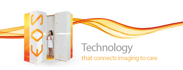
PARIS–(BUSINESS WIRE)–EOS imaging (Paris:EOSI) (Euronext, FR0011191766 – EOSI), the pioneer in 2D/3D orthopedic medical imaging, announced today the official opening of the first EOS site in South Korea, the third largest market in Asia, at the Konyang University Hospital. The University Hospital serves an adult and pediatric population of 7 million outpatients and 25 million inpatients on average per year.

The University Hospital has a strong orthopedics department for surgical and non-surgical treatment of the spine and joints. Patients of the specialized spine clinic will benefit from the low dose, full body images and weight-bearing, 3D analysis that EOS provides for deformative and degenerative spine conditions. In addition, the joint replacement clinic will use the EOS images and 3D torsion and alignment measurements to improve the planning of arthroplasty surgeries and control post-operative outcomes.

Prof Kim Sang Beom, Chief of the Spine Centre and Orthopedics Department and Board Member of the Korea Spine Surgeon Association, commented, “EOS is a real innovative technology with low dose biplanar 2D/3D radiography that’s approved by NECA. We are happy to be able to extend our services and care to our patients with this technology, including beginning to screen children and students utilizing the applicable Micro Dose option. The Konyang University Hospital uses the most advanced technology for better patient outcome. EOS will be the gold standard for musculoskeletal treatment in the near future.”

Marie Meynadier, CEO of EOS imaging, commented, “The Konyang University Hospital will be a strong reference center for the EOS platform in South Korea. Together with the Innovative Technology status obtained from the NECA Center for New Health Technology earlier this year, this will fuel adoption in the important South Korean market. We look forward to serving more Korean hospitals, healthcare professionals and patients with our low dose 2D/3D technology.”

For more information, please visit www.eos-imaging.com.

EOS imaging has been chosen to be included in the new EnterNext© PEA-PME 150 index, composed of 150 French companies and listed on Euronext and Alternext markets in Paris.

EOS imaging is listed on Compartment C of Euronext Paris
ISIN: FR0011191766 – Ticker: EOSI

About EOS imaging

EOS imaging designs, develops, and markets EOS®, an innovative medical imaging system dedicated to osteoarticular pathologies and orthopedics, as well as associated solutions. The Company is authorized to market in 51 countries, including the United States, Japan, China, and the European Union. The Group posted 2015 revenues of €21.8 million and employs 122 people. The Group is based in Paris and has five subsidiaries in Besançon (France), Cambridge (Massachusetts), Montreal (Canada), Frankfurt (Germany) and Singapore.

Contacts

EOS imaging
Anne Renevot
CFO
Ph: +33 (0)1 55 25 61 24
investors@eos-imaging.com
or
NewCap
Financial communication and investor relations
Valentine Brouchot
Ph: +33 (0)1 44 71 94 96
eosimaging@newcap.eu
or
The Ruth Group (US)
Press relations / Joanna Zimmerman
Ph: 646-536-7006
jzimmerman@theruthgroup.com

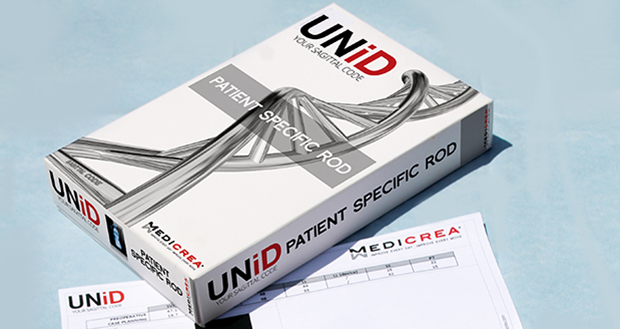
MEDICREA Achieves Personalized Spine Milestone with 1,000 Patient-Specific UNiD™ Rod Procedures

November 28, 2016

LYON, France & NEW YORK–(BUSINESS WIRE)–The MEDICREA Group (Alternext Paris: FR0004178572 – ALMED), worldwide leader pioneering the development and manufacture of personalized analytical services and implant solutions for the treatment of complex spinal conditions, announced today that the Company’s patient-specific UNiD™ Rod technology has now been used in more than 1,000 procedures.

Adoption of the UNiD™ technology continues to grow and gain momentum with over 110 orthopedic and neurosurgeons using UNiD™ Rods in the United States and Europe and represents a major milestone in establishing MEDICREA as an industry innovator in the global complex spine market. Surgeons discuss what UNiD™ means to their practice in this video. A special animation commemorating the 1,000-surgery milestone is also available here.

Dr. Evalina Burger, of University Colorado Hospital, stated, “We now realize how important it is to provide a specific alignment of the spine that is unique to each patient. If we do not achieve the optimal alignment during surgery, then we are too-often revising that patient later. UNiD™ is using precision technology and analysis to solve this clinical issue for patients as well as providing an invaluable support service and feedback loop for surgeons.” Dr. Burger has performed more than 40 procedures utilizing UNiD™ Rods since the patient-specific technology was FDA-cleared in November 2014 and published her early results in Orthopedics.

Denys Sournac, President and CEO, added, “It is MEDICREA’s unique vision to bring personalized planning and analytical services to complex spine, forming a virtuous cycle of clinical improvement. We believe UNiD™ technology has the potential to significantly improve patient treatment and limit the costly burden placed on healthcare providers by reducing the occurrence of revision surgery.”

The 1,000-procedure milestone is achieved on the heels of the Company’s UNiD™ LIFETIME WARRANTY launch, announced on October 25, 2016 with the UNiD™ Premier Service, making MEDICREA the only player in Spine to provide a guarantee against one of the leading causes of revision surgery in complex spinal indications: structural compromise to the rod.

MEDICREA’s UNiD™ Thoracolumbar and Cervical Rods provide a complementary solution to the Company’s comprehensive solution package for complex spine, including PASS LP® and PASS OCT® fixation systems. The systems are marketed by MEDICREA within a global marketspace representing an annual value estimated to near $6 billion for the thoracolumbar and cervical fixation segments.

MEDICREA anticipates the announcement of FDA 510k clearance in the coming weeks for the Company’s next extension to its complex spine portfolio: a range of top-loading, self-locking tulip screws, the most prevalent screw type in the complex spine market.

About MEDICREA (www.medicrea.com)

MEDICREA specializes in bringing pre-operative digital planning and pre and post-operative analytical services to the world of complex spine. Through the lens of predictive medicine, MEDICREA leads the design, integrated manufacture, and distribution of 30+ FDA approved implant technologies, utilized in over 100k spinal surgeries to date. Operating in a $10 billion marketplace, MEDICREA is an SME with 150 employees worldwide, which includes 55 at its USA Corp. subsidiary in NYC. The Company has an ultra-modern manufacturing facility in Lyon, France housing the development and production of 3D-printed titanium patient-specific implants.

By leveraging its proprietary software analysis tools with big data and deep learning technologies supported by an expansive collection of clinical and scientific data, MEDICREA is well-placed to streamline the efficiency of spinal care, reducing procedural complications and limiting time spent in the O.R.

For further information, please visit: medicrea.com.

Connect with MEDICREA:
FACEBOOK | INSTAGRAM | TWITTER | WEBSITE | YOUTUBE

MEDICREA is listed on ALTERNEXT Paris ISIN: FR 0004178572 – Ticker: ALMED

Contacts

MEDICREA
Denys Sournac
Founder, Chairman and CEO
dsournac@medicrea.com
or
Fabrice Kilfiger
Chief Financial Officer
fkilfiger@medicrea.com
+33 (0)4 72 01 87 87
or
Media
Russell Ward for MEDICREA
russell@theconfluencegroup.com
+1 310 424 8356

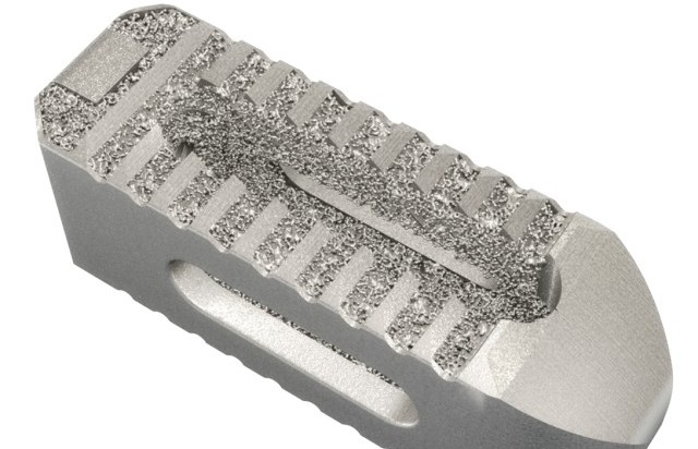
Stryker Conducts Successful Pre-Clinical Trial on 3D Printed Tritanium PL Interbody Cage

by | Nov 2, 2016

When it comes to utilizing 3D printing technology to produce medical devices and tools, there is usually a comprehensive trial that these printed materials must undergo before they can be applied in the healthcare sector. These pre-clinical studies help to unearth the benefits and disadvantages of these potential medical solutions, comparing them to other biocompatible materials that are commonly used in similar applications. The medial technology company Stryker is one of the pioneers of using 3D printing technology to enhance the production of medical devices and implants.

After announcing plans to start construction on a multimillion-dollar 3D printing manufacturing facility earlier this year, Stryker went on to develop their 3D printed Tritanium Posterior Lumbar (PL) Cage Spinal Implant, which was debuted at the American Association of Neurological Surgeons (AANS) Annual Scientific Meeting back in May. Last week, the Spine division of the medical technology company announced the completion of a pre-clinical study for this Tritanium PL Interbody Cage.

The results of the study compare their highly porous titanium alloy with the biomechanical, radiographic, and histological performance of commonly used implants with different surface technologies in an ovine lumbar interbody fusion model. The materials involved in the comparative study include traditional PEEK cages and plasma-sprayed titanium-coated PEEK cages. Using their proprietary Tritanium Technology, Stryker was able to produce a material with porous structures that resembles spongy bone tissue.


READ THE REST HERE

Spine Navigation Technology can ease surgery complications: Doctors

IANS | Nov 24, 2016

Spine Navigation Technology – an image-based medical technology – can be the best way to perform delicate and complex spinal surgeries, said doctors on Wednesday.

According to them, the biggest advantage of the Spine Navigation Technology is that doctors are able to operate with better visualisation and more accuracy than ever before.

“The image-based technology used in spinal surgery utilises scans of the patient’s anatomy and instruments that are tracked by the Navigation System’s camera. The specialised software creates a virtual, 3-D model of the patient’s spine, essentially a digital roadmap or blueprint to help guide the surgeon,” said Arvind Kulkarni, Head of Spine Scoliosis and Disc Replacement Centre at Bombay Hospital.

Adding further, he said: “The spine surgeon can use this model to plan the details of the surgery including the number, size and location of implants.”

He said that the new technology used during complex cases enables faster, precise and less invasive spinal procedures in a reduced radiation environment.


READ THE REST HERE

Clinical and retrieval evidence for corrosion of spine implants

All prosthetic implants are subject to some degree of wear and corrosion in the body. Wear is a mechanical process resulting from changes in load distribution and micro-motion, while corrosion is an electrochemical process of metal degradation. Certain types of corrosion, such as fretting, occur when chemical and mechanical factors, including crevice and abrasive wear, act on the implant.

Both wear and corrosion result in debris release from the implants into the surrounding tissues and subsequently into the human circulation, and can manifest with local and systemic reactions which, over time, may demand revision of components. Although numerous studies have focused on the clinical significance of corrosion and wear of hip and knee replacements, research involving spine instrumentation is not well documented.

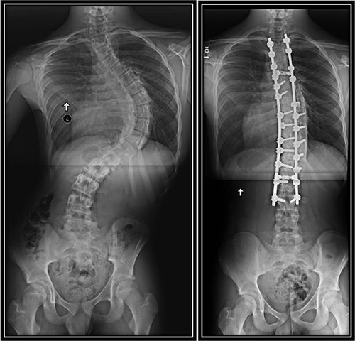
Recently, we received spine instrumentation, with cobalt–chrome (CoCr) rods with titanium (Ti) fixtures, of a 36-year-old female patient (Figure 1). She was diagnosed with adult idiopathic scoliosis (Figure 1A); however, soon after surgical implantation (Figure 1B) she experienced persistent pain and muscle fasciculation. Surgeons recommended removal of metalwork after two years of implantation, where intra-operatively they discovered metal staining of the periprosthetic tissue. We analysed the retrieved components in our facilities, and this case will serve here as a running example.


READ THE REST HERE

Minimally invasive lumbar decompression may be performed safely without prior experience

23rd November 2016

Junyoung Ahn (Department of Orthopedic Surgery, Rush University Medical Center, Chicago, USA) and others report in The Spine Journal that continued surgical experience in the context of minimally invasive lumbar decompression, with or without discectomy, is associated with reduced operative times and reduced hospital stay, but not improved clinical outcomes. They conclude that a minimally invasive lumbar decompression could, therefore, be performed safely without prior experience.

According to the authors, minimally invasive spinal surgery procedures “carry an inherently difficult” learning curve “based on anecdotal evidence”. Noting that few studies have investigated the learning curve associated with minimally invasive laminectomy or laminotomy, with or without discectomy, they sought to “describe the surgical curve” of this technique using a prospectively maintained surgical registry “with the largest surgical series by a single surgeon to date”.,,

In a primary analysis, the initial 50 consecutive patients of 228 who underwent a level one or two minimally invasive lumbar decompression (with or without discectomy) by a single surgeon were compared with the remaining 178. “Perioperative parameters (preoperative visual analogue scale scores, procedural, estimated blood loss, and length of hospital stay) were assessed between cohorts. Postoperative visual analogue scale (VAS) scores and complications were also compared between cohorts,” Ahn et al report.

Both procedural time (52.2±2.2 minutes versus 39.4±19.8 minutes; p<0.001) and length of hospital stay (22.1±25.4 hours versus 15.2±15 hours; p<0.01) were significantly reduced in the second cohort.

Furthermore, the authors comment: “Pearson correlation coefficient demonstrated that the case number is related to decreased surgical time (r=0.23; p<0.001) and decreased length of hospitalisation (r=0.12; p=0.08).” They add that their findings are consistent with those of a study that suggested the learning curve for minimally invasive lumbar discectomy was between 25 and 100 cases, commenting that—in the present study—the “procedural time plateaued following 50 cases, signifying the learning curve of the minimally invasive lumbar decompression”.

However, estimated blood loss, VAS scores, body mass index, complication rates, and recurrent herniation rates (index and adjacent levels) were similar between cohorts. “The rates of reoperation at the index level did not differ between the first and second cohorts (12% versus 8.4%, respectively).

No differences were demonstrated in the rates, indications, and the type of second surgery between cohorts. In addition, no differences were demonstrated in the reoperation rates before and after 12 months following the index procedure,” Ahn et al comment. According to the authors, the recurrent herniation rates, for both cohorts, were “within range” of those reported in previous studies.

Furthermore, in a secondary analysis, Ahn et al compared the first cohort with 50 patients who had undergone open lumbar decompression (by the same surgeon). They found that open surgery was associated with greater procedural time, higher estimated blood loss and longer length of hospital stay, but had similar reoperation rates and 30-day readmission rates to the minimally invasive surgery patients.

Concluding their findings, the authors comment that they “do believe that the refinement of the technique occurs with time as evidenced by the lower reoperation rate (not statistically significant) in the second cohort” but add—given the comparable outcomes and safety between cohorts—that minimally invasive lumbar decompression may initially be performed “without prior experience”. “We believe this finding may be helpful and comforting for those surgeons contemplating the incorporation of minimally invasive surgical decompression techniques into their practice,” Ahn et al add.

Co-author Kern Singh (Chicago, USA) told Spinal News International, “There are many factors that may possibly affect the learning curve for different surgeons. One potential factor includes the amount of prior experience a surgeon has in performing both open and minimally invasive procedures. While it is logical to assume that a more experienced surgeon will have a reduced learning curve for new procedures, further study is necessary to confirm that notion. Multi-surgeon studies are required to better elucidate the effect of surgeon training and years in practice on the learning curve for minimally invasive procedures.”

Writing in an accompanying commentary, Choll W Kim (San Diego, USA) notes that the learning curve “remains a significant factor” affecting the adoption of minimally invasive spinal surgery technologies. He adds that an important aspect of the learning curve “that is often neglected” is the specific training to the surgeon in the operating room before the first case, noting “little attention has been given to the training and preparations of the surgical team for a specific new procedure”. According to Kim, the “skin-to-skin” programme—an intensive two-day course on minimally invasive surgery—focuses on performing procedures multiple times and initial results indicate that it is associated with an improved learning curve and could lead to greater adoption of minimally invasive techniques.

“It is reasonable to assume that the learning curve is not static; it can be affected in multiple ways: training, preparation, changes in equipment, technique modifications, and even identification of disease states and patient factors that make certain surgical procedures more or less difficult. To do this, measurement parameters of the learning curve must be established,” he states.