Orthofix Announces First Patient Enrollment in Osteoarthritis of the Knee Study

September 12, 2016

LEWISVILLE, Texas–(BUSINESS WIRE)–Orthofix International N.V., (NASDAQ:OFIX), a diversified, global medical device company, today announced enrollment of the first patient in a study evaluating the use of pulsed electromagnetic field (PEMF) technology for osteoarthritis (OA) of the knee. This study will assess the efficacy and safety of the Company’s Physio-Stim system in reducing inflammation and restoring homeostasis of the extracellular matrix, potentially providing symptomatic relief of OA pain, reducing cartilage breakdown and stimulating new cartilage formation. If successful, this would be the first disease-modifying treatment for osteoarthritis. Preservation or restoration of joint structure together with reduction of inflammation would be expected to improve symptoms and reduce the need for surgical intervention.

The first patient in the study was enrolled by Dr. Roy Aaron, Professor at Brown University in Providence, RI. “Finding new solutions for OA of the knee is important as these patients not only suffer from pain, they often have a high level of disability from the disease and often seek costly surgical solutions,” said Dr. Aaron. “We are hopeful that active PEMF stimulation may provide us with a new non-invasive treatment approach that could help patients with knee OA avoid surgery and improve their overall quality of life.”

The OA of the knee study is a prospective, randomized, double-blind, placebo-controlled feasibility trial that will enroll approximately 150 patients who are 40 years of age or older at three sites in the U.S. These include Brown University School of Medicine in Providence, RI; Sinai Hospital of Baltimore in Baltimore, MD; and the Cleveland Clinic in Cleveland, OH. Study participants will be randomized in a two-to-one ratio to either an active or placebo control (inactive) device and followed for 12 months after initiation of treatment.

“This study demonstrates Orthofix’s vision of developing new applications for our PEMF technology,” said James Ryaby, Ph.D., Chief Scientific Officer for Orthofix. “We are proud to conduct research studies like these in order to find solutions for patients who live with disabling diseases and conditions such as OA.”

The Orthofix Physio-Stim device is currently approved by the U.S. Food and Drug Administration (FDA) for the treatment of nonunion fractures. The device uses a low-level electromagnetic field (PEMF) that helps activate and augment the body’s natural healing process. Use in the OA study is investigational and is being conducted under an Investigational Device Exemption (IDE) from the FDA. More information is available at ClinicalTrials.gov.

About Osteoarthritis of the Knee

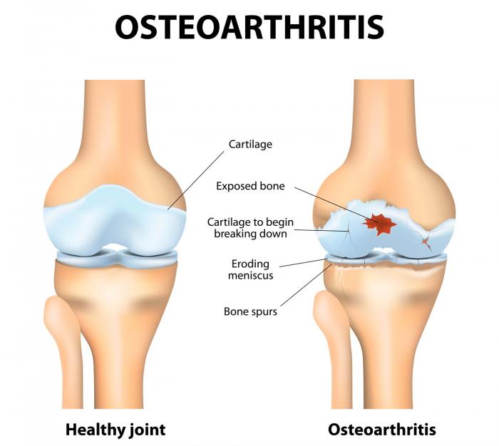
Osteoarthritis is a common disease and one of the leading causes of disability. The Centers for Disease Control and Prevention estimate that nearly one in two people may develop symptomatic knee OA by age 85. Statistics show that in 2011 alone, there were 757,000 total knee replacement procedures performed, with OA accounting for 95 percent of these surgeries. Osteoarthritis of the knee is a degenerative, “wear-and-tear” type of arthritis where the cartilage in the knee joint gradually wears away. This painful disease most often effects people 50 years of age and older, but may occur in younger people, too. People with OA of the knee often experience pain when walking, running, bending or kneeling. They may also experience joint stiffness and swelling, making it difficult to do many everyday activities.

About Orthofix

Orthofix International N.V. is a diversified, global medical device company focused on improving patients’ lives by providing superior reconstructive and regenerative orthopedic and spine solutions to physicians worldwide. Headquartered in Lewisville, TX, the company has four strategic business units that include BioStim, Biologics, Extremity Fixation and Spine Fixation. Orthofix products are widely distributed via the company’s sales representatives, distributors and subsidiaries. In addition, Orthofix is collaborating on research and development activities with leading clinical organizations such as Brown University, Sinai Hospital of Baltimore, Cleveland Clinic, Texas Scottish Rite Hospital for Children and the Musculoskeletal Transplant Foundation. For more information, please visit www.orthofix.com.

Forward-Looking Statements

This communication contains certain forward-looking statements under the Private Securities Litigation Reform Act of 1995. These forward-looking statements, which may include, but are not limited to, statements concerning the projections, financial condition, results of operations and businesses of Orthofix and its subsidiaries, are based on management’s current expectations and estimates and involve risks and uncertainties that could cause actual results or outcomes to differ materially from those contemplated by the forward-looking statements. The forward-looking statements in this release do not constitute guarantees or promises of future performance. Factors that could cause or contribute to such differences may include, but are not limited to the risks described in the “Risk Factors” section of our 2015 Annual Report on Form 10-K, as well as in other reports that we file in the future. Existing and prospective investors are cautioned not to place undue reliance on these forward-looking statements, which speak only as of the date hereof. The Company undertakes no obligation to update or revise the information contained in this press release.

Contacts

Orthofix International N.V.
Investor Relations
Mark Quick, 214-937-2924
markquick@orthofix.com
or
Media Relations
Denise Landry, 214-937-2529
deniselandry@orthofix.com

Intuitive Surgical Reports New Employee Option Grants for September 2016

SUNNYVALE, Calif., Sept. 09, 2016 (GLOBE NEWSWIRE) — Intuitive Surgical, Inc. (ISRG) today reported that equity awards approved by the Compensation Committee of the Board of Directors, which consists entirely of Independent Directors, were made to 86 new employees. Pursuant to NASDAQ Marketplace Rule 5635(c)(4), the equity awards were granted under the Intuitive Surgical, Inc. 2009 Commencement Incentive Plan, which the Board of Directors of Intuitive Surgical, Inc. adopted for the granting of equity awards to new employees. In accordance with NASDAQ rules, these grants were made under an equity incentive plan without shareholder approval. NASDAQ rules require a public announcement of equity awards to be made under this type of plan.  86 employees were granted a combination of Restricted Stock Units (RSUs) and Stock Options to purchase an aggregate of 10,245 shares of the Company’s common stock; 4,816 of the shares granted were Stock Options and 5,429 of the shares granted were RSUs. Both the RSUs and Stock Options vest over four years.  The Stock Options expire in 10 years assuming continued employment. No officers received any award under this plan. The exercise price for the Stock Options granted is $691.15 which was the closing price of Intuitive Surgical, Inc.’s common stock on the NASDAQ Global Market as such price was reported by NASDAQ on September 8, 2016. The Company’s policy is to issue RSUs and Stock Option grants to new employees, where equity makes sense, on the fifth business day of every calendar month.

About Intuitive Surgical, Inc.

Intuitive Surgical, Inc. (ISRG), headquartered in Sunnyvale, California, is the global technology leader in robotic-assisted, minimally invasive surgery. Intuitive Surgical develops, manufactures and markets robotic technologies designed to improve clinical outcomes and help patients return more quickly to active and productive lives. The Company’s mission is to extend the benefits of minimally invasive surgery to the broadest possible base of patients. Intuitive Surgical – Taking surgery beyond the limits of the human hand™.

About the da Vinci® Surgical System

The da Vinci® System is a breakthrough surgical platform designed to enable complex surgery using a minimally invasive approach. The da Vinci® System consists of an ergonomic surgeon console, a patient-side cart with four interactive robotic arms, a high-performance vision system and proprietary EndoWrist® instruments. Powered by state-of-the-art robotic and computer technology, the da Vinci® System is designed to scale, filter and seamlessly translate the surgeon’s hand movements into more precise movements of the EndoWrist® instruments. The net result is an intuitive interface with breakthrough surgical capabilities. By providing surgeons with superior visualization, enhanced dexterity, greater precision and ergonomic comfort, the da Vinci Surgical System makes it possible for more surgeons to perform minimally invasive procedures involving complex dissection or reconstruction. This ultimately has the potential to raise the standard of care for complex surgeries, translating into numerous potential patient benefits, including less pain, a shorter recovery and quicker return to normal daily activities.

Intuitive®, da Vinci®, da Vinci S®, da Vinci® Si™, InSite® and EndoWrist® are trademarks or registered trademarks of Intuitive Surgical, Inc.

For more information, please visit the company’s web site at www.intuitivesurgical.com.

Jefferson Receives $1 Million from Businessman and Philanthropist Jamie Maguire and the Maguire Foundation to Endow Professorship in Spine Research

(PHILADELPHIA) – Jamie Maguire, chairman of Philadelphia Insurance Companies (PHLY), and the Maguire Foundation, have generously funded a new orthopaedics professorship at Thomas Jefferson University.

The gift of $1 million will be matched dollar for dollar by the Sidney Kimmel Foundation Matching Program at Jefferson to establish the James J. Maguire, Jr. Endowed Professorship for Spine Research.

“We are incredibly grateful for this gift and for Jamie’s personal advocacy efforts related to health and wellness,” said Alexander R. Vaccaro, MD, PhD, MBA, the Richard H. Rothman Professor and Chair of Orthopaedic Surgery at Sidney Kimmel Medical College at Thomas Jefferson University and President of the Rothman Institute. “The Maguire family’s support will help us fulfill our mission to advance orthopaedic science and technology to benefit patients of all ages—in this case, specifically those with debilitating spinal conditions and injuries.”

Maguire’s passion for advancing orthopaedic research and clinical care stems from his personal experience recovering from a cycling accident. A longtime Ironman competitor, he was training for a race in 2014 when he crashed into a fallen tree and became paralyzed from the neck down. With four broken vertebrae in his neck, Maguire underwent emergency surgery with a team from the Rothman Institute at Jefferson. Three days later, he had regained movement and was recovering at home. Today, he has full range of motion in his neck is back to participating in endurance sport events, including Ironman.

“Giving back is a mantra that I’ve lived by all my life,” said Maguire. “Jefferson gave me a second chance at living and enjoying my life, and I wanted to do something to help them do the same for other patients in need.”

“The Spine program at Jefferson, under the leadership of Dr. Alex Vaccaro and the Rothman Institute, is internationally recognized for its excellence. But that pales in comparison to the level of impact that the clinical care and research can have on an individual’s life,” said Stephen K. Klasko, MD, MBA, president and CEO of Thomas Jefferson University and Jefferson Health. “Jamie Maguire and the Maguire Foundation’s generous investment will recognize the achievements of an exceptional faculty member while providing sustained resources for breakthrough treatments, diagnoses and new cures for years to come.”

In addition to his philanthropic support, Maguire serves as a Rothman Ambassador, sharing his story to help spread awareness about the research, care and compassion provided by the Rothman Institute at Jefferson. Watch his story here:https://www.rothmaninstitute.com/stories/ambassadors.

Media Contact
Gianna DeMedio
Gianna.DeMedio@Jefferson.edu
Phone: 215-955-5507

VTI Announces Partnership With BioMed. Tech. Intl. S.A. (BioMedical)™

MINNETONKA, MN (PRWEB) SEPTEMBER 09, 2016

VTI, Vertebral Technologies, Inc., announced a strategic partnership with BioMed. Tech. Intl. S.A. (BioMedical), the Mexico-based medical products distributor whose aim is to represent technology that helps people reclaim their lives.

BioMed Tech. Intl. will offer distribution of VTI’s patented InterFuse S™ and InterFuse T™ spinal fusion devices throughout Mexico.

BioMed Tech. Intl. President and CEO Marco Vega comments, “We are proud to work with the most important opinion leaders on MIS Spine Surgery in Mexico and Latin America. As a MIS Spine distributor leader we have the responsibility to increase the awareness of new and experienced physicians with the practice of MIS surgery”. Mr. Marco Vega also stated, “We decided to carry Interfuse in Mexico, because we are a Company who handles the most advanced Minimally Invasive Spine Products for our MIS Surgeons and Patients”.

The InterFuse™ System is a spinal implant able to achieve a large footprint through an intra-body modular assembly technique, using a minimally invasive posterior and or transformational approach. The InterFuse™ is currently being used by surgeons in over 20 countries, and VTI is pleased to be able to bring this state-of-the-art technology to more spine specialists in the country of Mexico.

“We have seen significant interest from Mexican surgeons at the various international congresses and we are looking forward to bringing the unique benefits of InterFuse™ to the Mexican Spine Surgeons and their Patients.” says VTI Vice President of International Sales, Ben Wasscher. He continues by stating, “Biomedical is a well-respected distributor in the Mexican spine market with great geographic coverage through its 6 offices and its large team of motivated sales people.”

This partnership will be formally announced at the AMCICO Spine Congress in Mexico City, September 13-17, 2016. This is the 17th Congress organized by AMCICO (Asociacion Mexicana de Cirujanos de Columna / Mexican Association of Spine Surgeons/ Spine Surgeons Association of Mexico), and both VTI and BioMed Tech. Intl. representatives will be in attendance.

About BioMedical Tech. Intl. (BioMedical).

Founded in 2004, BioMedical Tech. Intl., is a private held company who focuses on bringing high quality medical products and innovative technology to surgeons. Their Sales Operations Centers are located throughout Mexico, in Mexico City (headquarters), Guadalajara, Tijuana, Monterrey, Chihuahua, Merida, Villahermosa, Veracruz, Hermosillo and with a range of Subdistributors all around the country.. BioMedical’s mission is “To allow people to live more fully and healthier by seeking excellent quality in the products we manage, and to deliver unbeatable service at the appropriate time.” For more information for VTI products in Mexico, you can visit: http://http://www.biomedical.mx or contact Pedro Montero for sales at: +52 (55) 52638370.

About Vertebral Technologies, Inc.

Vertebral Technologies, Inc. (VTI) is a privately held company based in Minnetonka, MN, USA. VTI is dedicated to the design, development, manufacturing and marketing of medical devices to address painful conditions of the spine through less-invasive surgical approaches. VTI’s products utilize its unique modular-assembly technology to deliver solutions optimized for both surgeons and their patients. VTI sells its InterFuse modular interbody fusion devices worldwide to-date.

For more information visit, http://www.vti-spine.com or contact Brian Thron at marketing(at)vti-spine(dot)com or +1.952-979-1811

Chemists devise revolutionary 3D bone-scanning technique

Chemists from Trinity College Dublin, in collaboration with RCSI, have devised a revolutionary new scanning technique that produces extremely high-res 3D images of bones — without exposing patients to X-ray radiation.

The chemists attach luminescent compounds to tiny gold structures to form biologically safe ‘nanoagents’ that are attracted to calcium-rich surfaces, which appear when bones crack – even at a micro level. These nanoagents target and highlight the cracks formed in bones, allowing researchers to produce a complete 3D image of the damaged regions.

The technique will have major implications for the health sector as it can be used to diagnose bone strength and provide a detailed blueprint of the extent and precise positioning of any weakness or injury. Additionally, this knowledge should help prevent the need for bone implants in many cases, and act as an early-warning system for people at a high risk of degenerative bone diseases, such as osteoporosis.

The research, led by the Trinity College Dublin team of Professor of Chemistry, Thorri Gunnlaugsson, and Postdoctoral Researcher, Esther Surender, has just been published in the leading journal Chem, a sister journal to Cell, which is published by CellPress.

Professor Gunnlaugsson said: “This work is the outcome of many years of successful collaboration between chemists from Trinity and medical and engineering experts from RCSI. We have demonstrated that we can achieve a three-dimensional map of bone damage, showing the so-called microcracks, using non-invasive luminescence imaging. The nanoagent we have developed allows us to visualise the nature and the extent of the damage in a manner that wasn’t previously possible. This is a major step forward in our endeavour to develop targeted contrast agents for bone diagnostics for use in clinical applications.”

The work was funded by Science Foundation Ireland and by the Irish Research Council, and benefited from collaboration with scientists at RCSI (Royal College of Surgeons in Ireland), led by Professor of Anatomy, Clive Lee.

Professor Lee said: “Everyday activity loads our bones and causes microcracks to develop. These are normally repaired by a remodelling process, but, when microcracks develop faster, they can exceed the repair rate and so accumulate and weaken our bones. This occurs in athletes and leads to stress fractures. In elderly people with osteoporosis, microcracks accumulate because repair is compromised and lead to fragility fractures, most commonly in the hip, wrist and spine. Current X ray techniques can tell us about the quantity of bone present but they do not give much information about bone quality.”

He continued: “By using our new nanoagent to label microcracks and detecting them with magnetic resonance imaging (MRI), we hope to measure both bone quantity and quality and identify those at greatest risk of fracture and institute appropriate therapy. Diagnosing weak bones before they break should therefore reduce the need for operations and implants – prevention is better than cure.”

In addition to the unprecedented resolution of this imaging technique, another major step forward lies in it not exposing X-rays to patients. X-rays emit radiation and have, in some cases, been associated with an increased risk of cancer. The red emitting gold-based nanoagents used in this alternative technique are biologically safe – gold has been used safely by medics in a variety of ways in the body for some time.

Dr Esther Surender, Trinity, said: “These nanoagents have great potential for clinical application. Firstly, by using gold nanoparticles, we were able to lower the overall concentration of the agent that would have to be administered within the body, which is ideal from a clinical perspective. Secondly, by using what is called ‘two-photon excitation’ we were able to image bone structure using long wavelength excitation, which is not harmful or damaging to biological tissues.”

She added: “These nanoagents are similar to the contrast agents that are currently being utilised for MRI within the clinic, and hence have the potential to provide a novel means of medical bone diagnosis in the future. Specifically, by replacing the Europium with its sister ion Gadolinium, we can tune into the MRI activity of these nanoagents for future use alongside X-ray and computed tomography (CT) scans.”

Professor Gunnlaugsson and his research team are based in the Trinity Biomedical Sciences Institute (TBSI), which recently celebrated its 5-Year anniversary. Professor Gunnlaugsson presented his research at a symposium to mark the occasion, along with many other world-leaders in chemistry, immunology, bioengineering and cancer biology.

Article: Two-Photon Luminescent Bone Imaging Using Europium Nanoagents, Esther M. Surender, Steve Comby, Brenton L. Cavanagh, Orlaith Brennan, T. Clive Lee, Thorfinnur Gunnlaugsson, Chem, doi: 10.1016/j.chempr.2016.08.011, published online 8 September 2016.

Paralyzed Man Regains Use of Arms and Hands After Experimental Stem Cell Therapy at Keck Hospital of USC

LOS ANGELES, Sept. 7, 2016 /PRNewswire/ — Keck Medical Center of USC today announced that a team of doctors became the first in California to inject an experimental treatment made from stem cells, AST-OPC1, into the damaged cervical spine of a recently paralyzed 21-year-old man as part of a multi-center clinical trial.

On March 6, just shy of his 21st birthday, Kristopher (Kris) Boesen of Bakersfield suffered a traumatic injury to his cervical spine when his car fishtailed on a wet road, hit a tree and slammed into a telephone pole.

Parents Rodney and Annette Boesen were warned there was a good chance their son would be permanently paralyzed from the neck down. However, they also learned that Kris could possibly qualify for a clinical study that might help.

Leading the surgical team and working in collaboration with Rancho Los Amigos National Rehabilitation Center and Keck Medicine of USC, Charles Liu, MD, PhD, director of the USC Neurorestoration Center, injected an experimental dose of 10 million AST-OPC1 cells directly into Kris’ cervical spinal cord in early April.

“Typically, spinal cord injury patients undergo surgery that stabilizes the spine but generally does very little to restore motor or sensory function,” explains Liu. “With this study, we are testing a procedure that may improve neurological function, which could mean the difference between being permanently paralyzed and being able to use one’s arms and hands. Restoring that level of function could significantly improve the daily lives of patients with severe spinal injuries.”

Two weeks after surgery, Kris began to show signs of improvement. Three months later, he’s able to feed himself, use his cell phone, write his name, operate a motorized wheelchair and hug his friends and family. Improved sensation and movement in both arms and hands also makes it easier for Kris to care for himself, and to envision a life lived more independently.

“As of 90 days post-treatment, Kris has gained significant improvement in his motor function, up to two spinal cord levels,” said Dr. Liu.  “In Kris’ case, two spinal cord levels means the difference between using your hands to brush your teeth, operate a computer or do other things you wouldn’t otherwise be able to do, so having this level of functional independence cannot be overstated.”

Doctors are careful not to predict Kris’ future progress.

“All I’ve wanted from the beginning was a fighting chance,” said Kris, who has a passion for fixing up and driving sports cars and was studying to become a life insurance broker at the time of the accident.  “But if there’s a chance for me to walk again, then heck yeah! I want to do anything possible to do that.”

Qualifying for the Study

Because the window for performing the surgery was tight, everything needed to go according to schedule in order for Kris to qualify.

Once Kris made the decision to pursue enrollment in the study, dozens of doctors, nurses, rehabilitation specialists and others sprang into action. Because he would need to provide voice confirmation of his desire to participate in the study, Kris had to be able to breathe without a ventilator. Weaning a patient from assisted breathing generally is a three-week process. He did it in five days with the help of a respiratory care team. He signed the paperwork and began a week of assessments, scans and other pre-surgery tests.

In early April, a surgical team from Keck Hospital of USC carefully injected 10 million AST-OPC1 cells directly into Kris’ cervical spine. Nearly six weeks later, Kris was discharged and returned to Bakersfield to continue his rehabilitation. Doctors reviewed his progress at seven days, 30 days, 60 days and 90 days post-injection, and Kris can look forward to detailed assessments after 180 days, 270 days and one year.

Rodney and Annette Boesen say they are amazed at the level of collaboration and cooperation that enabled their son to participate in the study. “So many things had to happen, and there were so many things that could have put up a roadblock,” marvels Rodney. “The people at Keck Medical Center of USC and elsewhere moved heaven and earth to get things done. There was never a moment through all of this when we didn’t think our son was getting world class care.”

About the Study

The pioneering surgery is the latest example of how the emerging fields of neurorestoration and regenerative medicine may have the potential to improve the lives of thousands of patients who have suffered a severe spinal cord injury.

The stem cell procedure Kris received is part of a Phase 1/2a clinical trial that is evaluating the safety and efficacy of escalating doses of AST-OPC1 cells developed by Fremont, California-based Asterias Biotherapeutics. AST-OPC1 cells are made from embryonic stem cells by carefully converting them into oligodendrocyte progenitor cells (OPCs), which are cells found in the brain and spinal cord that support the healthy functioning of nerve cells. In previous laboratory studies, AST-OPC1 was shown to produce neurotrophic factors, stimulate vascularization and induce remyelination of denuded axons. All are critical factors in the survival, regrowth and conduction of nerve impulses through axons at the injury site, according to Edward D. Wirth III, MD, PhD, chief medical director of Asterias and lead investigator of the study, dubbed “SCiStar.”

“At the 10 million cell level, we’re now in a dose range that is the human equivalent of where we were when we saw efficacy in pre-clinical studies,” says Wirth. “While we continue to evaluate safety first and foremost, we are also now looking at how well treatment might help restore movement in these patients.”

To qualify for the clinical trial, enrollees must be between the age of 18 and 69, and their condition must be stable enough to receive an injection of AST-OPC1 between the fourteenth and thirtieth days following injury.

Keck is one of six sites in the U.S. authorized to enroll subjects and administer the clinical trial dosage.

The first early data results from a group of patients in this study will become available on September 14, 2016.

Photography and video assets can be downloaded at http://www.keckmedicine.org/stem-cell-patient-media-assets/

For more information, go to www.keckmedicine.org/beyond

Logo – http://photos.prnewswire.com/prnh/20160120/324215LOGO


SOURCE Keck Medicine of USC

Related Links

http://www.keckmedicine.org/

Global Market Study on Cervical Total Disc Replacement Device: Growth in Cervical Degenerative Diseases, a Catalyst for CTDR Devices Market

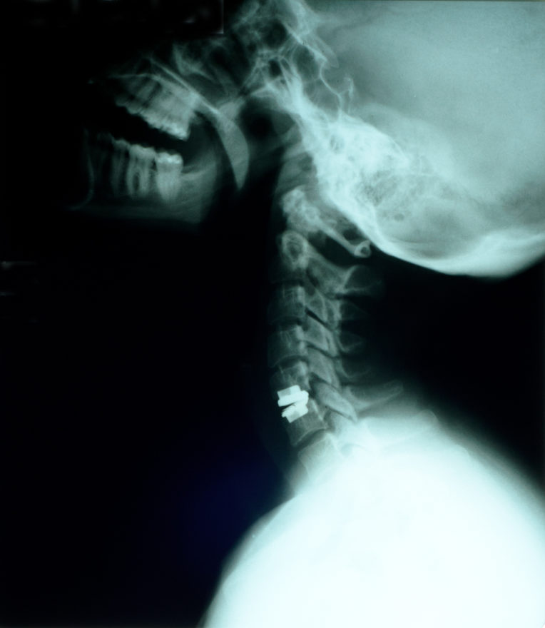
LONDON, Sept. 8, 2016 /PRNewswire/ — The cervical Total Disc Replacement (CTDR) device is used to treat patients suffering from Degenerative Disc Disorders (DDD). Neck pain is the major cause for the development of DDD in the cervical region of the spine. Cervical disc replacement surgery is performed for treatment of DDD, where the degenerated disc is removed surgically and an artificial disc is implanted in the vertebra. This is a Minimal Incision Surgery (MIS) and results in quick recovery post-surgery, short hospital stay, and low revision surgery rate.

Conventionally, DDD is treated through spinal fusion surgery, but due to the high rate of revision surgery and increasing the prevalence of adjacent segment disc degeneration post fusion surgery, doctors prefer cervical disc replacement over spinal fusion. Major players in the market are proactively providing training to spine surgeons, thus, ensuring efficient handling of their respective products.

Growing ageing population, low revision surgery rate, and increasing demand for advanced innovative CTDR devices for treatment of both class I and class II degenerative disc diseases, are major factors fueling demand for CTDR devices. Major trends witnessed in the CTDR device market include technological advancements, consistent investment in research and development activities, the and the surge in acquisitions and mergers.

The CTDR device market is segmented as follows:
CTDR device, by material type
CTDR device, by end user
CTDR device, by region
This report covers the global CTDR device market performance in terms of revenue contribution. This section also includes PMR’s analysis of key trends, drivers, restraints, and opportunities, which are influencing the growth of the global CTDR device market currently. Impact analysis of key drivers and restraints is included in this report to better equip clients with crystal clear decision-making insights. The report also provides regional company share analysis and product details in terms of marketing approvals.

By material type, the global CTDR device market is segmented into metal on a biocompatible material (M-o-B) and metal on metal (M-o-M). Of these, metal on biocompatible material segment holds majority market share currently and is expected to remain the leading segment in terms of volume and value during the forecast period. The metal on a metal segment is projected to grow in terms of value and volume, but at a relatively slower pace compared to the metal on the biocompatible material segment. The number of products in the metal on the biocompatible material segment is higher compared to the metal on a metal segment, which is a major driver for the growth of the metal on the biocompatible material segment. Various benefits offered by metal on biocompatible material products such as greater degree of rotation and flexible movement of a spine are expected to drive the growth of this segment during the forecast period.

On the basis of the end user, the global CTDR device market is segmented into hospitals and ASCs. The hospitals segment dominated the CTDR device market in 2014; however, the ASCs segment is projected to expand at a higher CAGR over the forecast period as compared to the hospital segment. This is attributed to the fact that cervical disc replacement is a minimally invasive surgery and can be performed in ASCs. Number of patients undergoing cervical disc replacement in ASCs is increasing substantially due to factors such as quick discharge and their cost effectiveness, which in turn is expected to fuel growth of CTDR device market in the ASCs segment.

By region, North America is expected to remain dominant in the CTDR device market mainly due to robust reimbursement policies and rise in the number of cervical disc replacement procedures performed in the region. Prevalence of neck pain is comparatively higher in Western Countries as compared to APAC. In terms of growth rate, the APAC market is projected to grow at a significant pace mainly due to the large population base in the region. Revenue contribution from the market in North America, followed by markets in APAC and Europe, is expected to expand at the highest CAGR during the forecast period. The market in the MEA remains untapped, thus offering potential opportunities to companies operating in this industry.

Some key market players included in this report are Medtronic, plc, Depuy Synthes (a Johnson & Johnson Company), Globus Medical, Inc., NuVasive, Inc., and LDR Holding Corporation. These companies have been profiled on the basis of various attributes such as company overview, recent developments, growth strategies, and financial overview.
Download the full report: https://www.reportbuyer.com/product/3403273/

About Reportbuyer
Reportbuyer is a leading industry intelligence solution that provides all market research reports from top publishers
http://www.reportbuyer.com

For more information:
Sarah Smith
Research Advisor at Reportbuyer.com
Email: query@reportbuyer.com
Tel: +44 208 816 85 48
Website: www.reportbuyer.com

SOURCE ReportBuyer

Related Links

http://www.reportbuyer.com

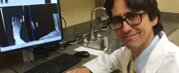
New foot and ankle surgeon joins Coastal Orthopedics

September 7, 2016

BRADENTON, Fla. — Dr. Andrés E. O’Daly, an orthopedic surgeon specializing in foot and ankle treatments, has just joined the comprehensive and full-service practice Coastal Orthopedics Sports Medicine & Pain Management (CoastalOrthopedics.com).

For the past year, Dr. O’Daly has been completing a foot and ankle surgery fellowship through the Department of Orthopaedic Surgery at the University of Alabama at Birmingham. He earned his Doctor of Medicine degree and completed his first orthopedic residency at the Central University of Venezuela.

“Since I was in Venezuela, I was interested in the mechanics of the foot,” said Dr. O’Daly. “It’s a growing, constantly evolving specialty with lots of innovation.”

Dr. O’Daly practiced as an orthopedic surgeon in Centro Medico de Caracas, one of the most prestigious clinics in Venezuela, focusing on sports medicine and joint reconstruction.

While receiving his medical training, Dr. O’Daly volunteered for a two-year clinical program at the Venezuelan Red Cross and interned at the Dr. Raúl Perdomo Hurtado Naval Hospital in Venezuela.

Dr. O’Daly earned his second residency from Johns Hopkins University after completing a research fellowship on peripheral nerve regeneration.

At Coastal Orthopedics, Dr. O’Daly will treat patients suffering from a variety of foot and ankle issues, including athletic injuries, fractures, dislocations, sprains, heel pain, Achilles tendon problems, arthritis and more.

“At Coastal Orthopedics, the motive is always patient care first,” said Dr. O’Daly. “I love the people at Coastal Orthopedics and the office.”

In the coming months, Dr. O’Daly will continue to be involved in an ongoing research project in conjunction with Johns Hopkins and the University of Alabama at Birmingham focused on ankle joint fracture care.

When he was working toward his fellowship at Johns Hopkins, Dr. O’Daly was honored with the Frank L. Coulson Jr. award for Clinical Excellence for several accomplishments, including communication and interpersonal skills, professionalism and humanism, diagnostic acumen, skillful negotiation of the healthcare system, a scholarly approach to clinical practice and a passion for patient care.

Dr. O’Daly, who recently moved to Bradenton with his wife and two daughters, is a member of the American Association of Orthopaedic Surgery and the American Orthopaedic Foot and Ankle Society.

About Coastal Orthopedics Sports Medicine & Pain Management
Founded in 1973 and based in Bradenton, Fla., Coastal Orthopedics is home to 15 orthopedic surgeons and pain management physicians who maintain hospital affiliations with Blake Medical Center, Manatee Memorial Hospital and Lakewood Ranch Medical Center. In addition to three offices and two ambulatory surgery centers each serving Florida’s West Coast, Coastal Orthopedics’ board-certified and fellowship-trained physicians and surgeons provide patients with the very latest in pain and orthopedic treatment technology. The company strives to repair and heal injuries relating to the entire musculoskeletal system. For more than 40 years, our highly skilled staff of physicians and surgeons has treated patients in specialty care including sports medicine, joint replacement, pain management and rehabilitation. Patients can experience restoration of function both surgically and non-surgically through the use of physician-guided strengthening, therapy, medications, injections and stretching. For more information, please visit CoastalOrthopedics.com or call 941-792-1404.


Carestream’s OnSight 3D Extremity System Receives FDA 510(k) Clearance

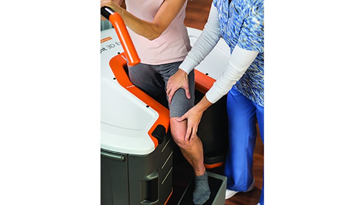
ROCHESTER, N.Y.–(BUSINESS WIRE)–The CARESTREAM OnSight 3D Extremity System that uses cone beam CT (CBCT) technology to capture weight-bearing and other types of extremity exams has received FDA 510(k) clearance and is available for order in the United States.

This affordable, compact system offers high-quality, lower-dose 3D imaging studies for use by orthopaedic and sports medicine practices, hospitals, imaging centers, urgent care facilities and other healthcare providers.

“Our extremity imaging system can help in treating a host of orthopaedic conditions that affect the biomechanical behavior of the joints such as arthritis, meniscus loss, instability and malalignment syndromes,” said Helen Titus, Carestream’s Worldwide Marketing Director for Ultrasound & CT Solutions. “This new system offers less radiation than traditional CT systems while delivering excellent image quality.”

Orthopaedic imaging is a major focus for Carestream because of the prevalence of musculoskeletal conditions among people of all ages. “Youth and adults often suffer sports-related injuries to their knees, ankles and feet while older adults experience arthritis, joint instability, meniscus loss and other conditions,” Titus explains.

Carestream’s new extremity imaging system enables healthcare providers to capture high-quality 3D images and conduct a patient consultation in a single visit—which helps improve productivity and convenience for both specialists and patients. An additional benefit is the ability for patients to view a 3D image that illustrates their condition or injury to help them understand the reason for a treatment or surgical procedure.

This new imaging system will be demonstrated at the American Society for Surgery of the Hand (ASSH) conference that begins on Sept. 29.

About Carestream Health

Carestream is a worldwide provider of dental and medical imaging systems and IT solutions; X-ray imaging systems for non-destructive testing; and precision contract coating services for a wide range of industrial, medical, electronic and other applications—all backed by a global service and support network. For more information about the company’s broad portfolio of products, solutions and services, please contact your Carestream representative or call 888-777-2072 or visit www.carestream.com. To view Carestream’s latest news announcements, please visit www.carestream.com/news.

CARESTREAM is a trademark of Carestream Health.

Follow Carestream Health online:

http://www.twitter.com/carestream
http://www.youtube.com/carestream
http://www.carestream.com/blog/
http://www.facebook.com/carestream
http://www.linkedin.com/company/carestream-health

Contacts

Carestream Health
Robert Salmon, 585-627-6560
robert.salmon@carestream.com

EOS imaging Reports First-half 2016 Results

September 08, 2016 12:29 PM Eastern Daylight Time

PARIS–(BUSINESS WIRE)–EOS imaging (Paris:EOSI) (Euronext, FR0011191766 – EOSI – Eligible for PEA-PME savings schemes in France), the pioneer in 2D/3D orthopedic medical imaging, announced today consolidated results for the six months ended June 30, 2016, as approved by the Board of Directors on September 8, 2016.

In millions of euros   First-half 2016   First-half 2015
   
Operating income
Revenue 14,1 10,2
Other income   1,17   0,96
Total income   15,3   11,2
Operating expenses
Direct cost of sales   -7,64   -5,19
Gross margin 6,51 5,01
as a % of revenues   46,0%   49,1%
Indirect cost of production and services -1,71 -1,47
Research & development -1,78 -1,71
Sales & marketing -3,62 -3,08
Regulatory expenses -0,35 -0,36
Administrative costs   -1,70   -1,74
Total operating expenses (excluding direct cost of sales and share-based payments)   -9,17   -8,36
Share-based payments   -0,24   -0,11
Total operating expenses   -17,04   -13,66
Net operating income/(loss)   -1,73   -2,51
Financial Income   -0,67   -0,11
Net income/(loss)   -2,40   -2,62
 
  • Strong sales growth in the first half of 2016: up 39% to €14.1 million

The Company sold 28 EOS® systems during the first half of the year, as compared to 20 during the same period last year. Revenues from sales of equipment totalled €11.45 million, an increase of 35%.

Sales of maintenance contracts increased 68% to €2.22 million, reflecting the continued increase in the installed base of EOS systems under contract.

Sales of consumables and services were €0.46 million in the first half of 2016, an increase of 23%.

The Company also recorded €1.17 million in other income consisting of public financing in support of its innovation (research tax credit and subsidies).

  • Gross margin: €6.5 million representing 46% of revenues

Gross margin for the first half of 2016 was 46% of revenues, as compared to 49% during the same period last year. This reflects a lower average selling price of equipment as compared to the first half of 2015.

  • Controlled increase in operating expenses

Operating expenses, excluding the direct cost of sales and share-based payments, were €9.17 million in the first half of 2016, up from €8.36 million in the first half of the previous year, representing an increase of 10%. This compares favorably to the 39% growth in revenue during the first half of 2016.

Sales & marketing expenses increased 18% in the first half of 2016, reflecting increased resources to support business growth. The 16% increase in indirect costs of production and services reflects the investments in support functions (quality and supply chain). The trend for other operating expenses (-3% to +4%) reflects controlled utilization of resources to support business growth.

Including the impact of share-based payments, the Company’s operating loss was €1.73 million in the first half of 2016, representing a reduction of the operating loss of 31% compared to an operating loss of €2.51 million in the same period last year.

The Company’s net loss for the six months ended June 30, 2016 was €2.40 million, compared with €2.62 million in the same period of 2015.

The Company had 128 employees as of June 30, 2016, compared with 122 employees as of December 31, 2015.

  • Cash position and balance sheet

The Company’s net cash position at June 30, 2016 was €14.4 million, compared with €14.1 million at December 31, 2015. Cash burn from operations was €3.6 million, with the increase in the working capital requirement limited to €2.1 million. A third and last €5.0 million tranche of convertible bonds was subscribed in June 2016 to support the execution of growth.

Equity at June 30, 2016 was €25.5 million compared with €27.8 million at December 31, 2015 as a result of the loss posted for the first half of 2016.

  • Half-year financial report

EOS imaging’s half-year financial report can be downloaded from its web site at www.eos-imaging.com by selecting Documentation, then Financial reports from the Investors menu.

Marie Meynadier, CEO of EOS imaging, commented: “The first half of 2016 was highlighted by excellent sales momentum and the establishment of strategic and technology partnerships that we expect to drive our business. We are executing on our growth and innovation strategy while maintaining a strong focus on cost control, which is reflected in the significant improvement in our half-year financial results. We are committed to continuing in this direction as we work to position the EOS platform as a standard of care that combines accurate and low-dose imaging and patient-specific surgical planning for a growing number of patients around the world.”

For more information, please visit www.eos-imaging.com.

About EOS imaging

EOS imaging designs, develops, and markets EOS®, an innovative medical imaging system dedicated to osteoarticular pathologies and orthopaedics, as well as associated solutions. The Company is authorized to market in 51 countries, including the United States (FDA), Japan, China and the European Union (EC). The Group posted 2015 revenues of €21.8 million and employs 122 people. The Group is based in Paris and has five subsidiaries in Besançon (France), Cambridge (Massachusetts), Montreal (Canada), Frankfurt (Germany) and Singapore.

EOS imaging has been chosen to be included in the new EnterNext© PEA-PME 150 index, composed of 150 French companies and listed on Euronext and Alternext markets in Paris.

EOS imaging is listed on Compartment C of Euronext Paris
ISIN: FR0011191766 – Ticker: EOSI

Next press release: 2016 Third-quarter sales: Thursday October 27, 2016 (after market close)

Contacts

EOS imaging
Anne Renevot, +33 (0)1 55 25 61 24
CFO
investors@eos-imaging.com
or
NewCap
Communication Financière et Relations investisseurs
Valentine Brouchot, +33 (0)1 44 71 94 96
eosimaging@newcap.eu
or
Relations presse
Annie-Florence Loyer, +33 (0)1 44 71 00 12/ +33 (6) 88 20 35 59
afloyer@newcap.fr
or
Daphné Boccara, +33 (0)1 44 71 94 93
dboccara@newcap.fr
or
The Ruth Group (US)
Relations presse
Joanna Zimmerman, 646-536-7006
jzimmerman@theruthgroup.com