Novastep Enhances its Nexis® Headless Screws Portfolio

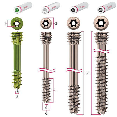
Orangeburg, New York, Oct. 07, 2016 (GLOBE NEWSWIRE) — Novastep, Inc., and its affiliates (“Novastep” or the “Company”), a global medical technologies company specializing in the foot and ankle, has further expanded its coverage of midfoot and rearfoot indications with the addition of a full range of Nexis® 5.0 and 7.0mm headless, cannulated compression screws.

The implants feature all of the hallmark design attributes inherent to the Company’s Nexis® brand, including helical thread flutes that are self-drilling, self-tapping and reverse-cutting; Torx drive recess interfaces for optimal force distribution and tapered, self-penetrating compression cones.  Loïc Girod, Novastep’s Vice President of Research and Development, noted that “The Nexis® system provides surgeons with a complete and versatile selection of sterile packaged, low-profile, cannulated screws that are designed to address a broad spectrum of midfoot and hindfoot pathologies.”

The system is likewise supported with a robust instrumentation platform that is neatly stored in a versatile, light-weight, space-saving tray to combine the Nexis® 5.0 and 7.0mm instruments with interchangeable Nexis® 4.0mm headless screw or Arcad 18/20/25mm nitinol compression clip instrument modules that serve to further enhance its versatility.  “Operating room efficiency and cost reduction are prime areas of focus for Novastep” said Joseph Larsen, DPM of ProHealthCare Foot & Ankle in New York; adding that “Novastep’s implant and cleanSTART® deployment technologies provide a systematized logistics platform that is easily customizable to fit virtually anyone’s needs and substantially reduces inventory requirements and sterile field volumes.”

All Novastep implants are packaged sterile in quickSTACK™ containers or quickTUBE™ nested cylinders, depending on the size and configuration of the product. The STACKS and TUBES are housed and organized in the cleanSTART® Implant Dispenser console, which may be uniquely tailored to address individual surgeon preferences.

Nexis® and other key elements of Novastep’s portfolio will be featured at the Desert Foot 2016 meeting on October 19–22 in Phoenix, AZ and the upcoming 2016 Global Foot and Ankle Symposium in New York City on December 2–3.

About Novastep

Novastep is a global medical device company specializing in the design, development and commercialization of advanced technologies that treat conditions affecting the foot and ankle. The Company is focused on optimizing clinical efficiencies, inventory management and healthcare economics by transforming the way foot and ankle products are deployed and utilized in the surgical environment. Novastep has allied itself with a strategic network of key international opinion leaders to deliver breakthrough technologies, innovative services and compelling medical education programs to the foot and ankle community. Novastep’s portfolio, services and distribution platforms are uniquely positioned to address foot and ankle trauma, deformity corrections and Charcot fracture management.

For further information concerning this announcement and/or Novastep, Inc., send all inquiries to info@novasteportho.com or call 877.287.0795.

Related Links

For general information visit:  novastep-us.com

EOS imaging Announces the Opening of a New EOS Installation at the Prestigious Charité Hospital in Berlin

October 10, 2016

PARIS–(BUSINESS WIRE)–EOS imaging (Paris:EOSI) (Euronext, FR0011191766 – EOSI), the pioneer in 2D/3D orthopedic medical imaging, announced today that the hospital Charité – Universitätsmedizin Berlin in Germany has started to provide patient exams with its new EOS® imaging system. Charité is a leading hospital in Germany with 17 different Charité Centers in the Berlin area.

Established over 300 years ago, Charité is one of the largest university hospitals in Europe. It employs 13,200 employees and handles 142,000 inpatient and 663,000 outpatient cases every year. A leading research institution, Charité lays claim to more than half of all German Nobel Prize winners in Physiology or Medicine.

Professor Dr. Carsten Perka, Medical Director of the Center for Musculoskeletal Surgery at Charité, commented, “The newly-acquired EOS system provides very accurate 3D imaging data for treatment planning. Of particular importance is that the EOS system provides a full-body exam in a functional position while conventional imaging examination positions are often unsuitable for evaluating the musculoskeletal system.”

Marie Meynadier, CEO of EOS imaging, said, “Charité is an internationally-recognized institution with a history of providing excellent care. The EOS installation there, adding to earlier installations in key private centers of the country, speaks to the growing recognition in Germany of our technology.”

For more information, please visit www.eos-imaging.com.

EOS imaging has been chosen to be included in the new EnterNext© PEA-PME 150 index, composed of 150 French companies and listed on Euronext and Alternext markets in Paris.

EOS imaging is listed on Compartment C of Euronext Paris
ISIN: FR0011191766 – Ticker: EOSI

About EOS imaging

EOS imaging designs, develops, and markets EOS®, an innovative medical imaging system dedicated to osteoarticular pathologies and orthopedics, as well as associated solutions. The Company is authorized to market in 51 countries, including the United States, Japan, China, and the European Union. The Group posted 2015 revenues of €21.8 million and employs 122 people. The Group is based in Paris and has five subsidiaries in Besançon (France), Cambridge (Massachusetts), Montreal (Canada), Frankfurt (Germany) and Singapore.

Contacts

EOS imaging
Anne Renevot
CFO
+33 (0)1 55 25 61 24
investors@eos-imaging.com
or
NewCap
Financial communication and investor relations
Valentine Brouchot
+33 (0)1 44 71 94 96
eosimaging@newcap.eu
or
The Ruth Group (US)
Press relations / Joanna Zimmerman
646-536-7006
jzimmerman@theruthgroup.com

Nanoparticle injections may be future of osteoarthritis treatment

By Jim Dryden

Osteoarthritis is a debilitating condition that affects at least 27 million people in the United States, and at least 12 percent of osteoarthritis cases stem from earlier injuries. Over-the-counter painkillers, such as anti-inflammatory drugs, help reduce pain but do not stop unrelenting cartilage destruction. Consequently, pain related to the condition only gets worse.

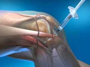
Now, researchers at Washington University School of Medicine in St. Louis have shown in mice that they can inject nanoparticles into an injured joint and suppress inflammation immediately following an injury, reducing the destruction of cartilage.

The findings are reported online Sept. 26 in the early edition of the Proceedings of the National Academy of Sciences.

“I see a lot of patients with osteoarthritis, and there’s really no treatment,” said senior author Christine Pham, MD, an associate professor of medicine. “We try to treat their symptoms, but even when we inject steroids into an arthritic joint, the drug only remains for up to a few hours, and then it’s cleared. These nanoparticles remain in the joint longer and help prevent cartilage degeneration.”

Frequently, an osteoarthritis patient has suffered an earlier injury — a torn meniscus or ACL injury in the knee, a fall, car accident or other trauma. The body naturally responds to such injuries in the joints with robust inflammation. Patients typically take drugs such as acetaminophen and ibuprofen, and as pain gets worse, injections of steroids also can provide pain relief, but their effects are short-lived.


READ THE REST HERE

New Extremity Products are Driving Growth for Arthrosurface

FRANKLIN, Mass., Oct. 6, 2016 /PRNewswire/ — Arthrosurface® announced today that at the September 27, 2016 American Society for Podiatric Surgery (ASPS) meeting in Baltimore, Maryland, the new CheckMATE® 3.0 Fusion Plate was launched for end stage toe arthritis. One study showed that 35%-60% of the population over 65 years old has osteoarthritis of the big toe, making this a common problem. The CheckMATE® plate was designed to offer advantages over existing systems by including features to make the plate low-profile, yet strong, rigid, and faster to implant, reducing surgical time and improving patient comfort. These features may also provide cost savings to the healthcare system and other clinical benefits to the patient. Arthrosurface is advancing patient care in the extremities market with a special focus in foot and ankle applications. Using evidence based designs that reference scientific literature, combined with clinical input from expert surgeon design teams, the Arthrosurface® extremities portfolio has expanded significantly over the past several years to provide innovative options for both surgeons and patients.

The CheckMATE® 3.0 complements the Arthrosurface® forefoot portfolio allowing surgeons to address the full spectrum of forefoot arthritis. With rising costs associated with valuable operating room time, shorter procedures become increasingly important. Various speed elements were built into the plate to reduce implantation time while still maintaining a state of the art forefoot fusion surgery. The CheckMATE® 3.0 plate is designed to minimize post-operative irritation and the need for secondary surgery for hardware removal, again providing a significant cost savings to the system and a potential improvement in patient comfort.

Dr. Patrick DeHeer, surgeon pioneer for the CheckMATE® plate commented, “The goal was to design a plate that is low-profile, but strong and rigid, providing the best of both internal and external fixation. This could allow patients to bear weight on their forefoot earlier, and to minimize secondary surgery and hardware irritation. This new plate has incorporated all these elements, making it a very attractive option for surgeons who perform first MTP fusions.”

“From a design perspective, our goal is to provide surgeons with complete and innovative solutions that improve upon the standard of care for patients experiencing forefoot arthritis. We want to challenge the status quo and use insights from the clinical data and our developers to make products that are anatomic, joint preserving and quick to implant,” said Steve Ek, Founder & CEO. “Arthrosurface is focused on bringing new products to the market that may enhance patient outcomes and potentially provide better options for both surgeons and patients in the extremities market,” commented Joseph Darling, Executive Chairman at Arthrosurface, Inc.

About Arthrosurface
Arthrosurface, Inc. is a leader in the design and distribution of orthopedic devices for joint preservation, restoration and resurfacing. The HemiCAP® system is a unique, less invasive technology that can be used to treat a wide variety of joint conditions caused by trauma, injury and disease. Founded in 2002, Arthrosurface markets and distributes its products in the US and around the world.

Logo – http://photos.prnewswire.com/prnh/20131118/NE19554LOGO

SOURCE Arthrosurface, Inc.

Related Links

http://arthrosurface.com

Flexion Therapeutics Announces Appointment of John Magee as Vice President of Sales

BURLINGTON, Mass., Sept. 29, 2016 (GLOBE NEWSWIRE) — Flexion Therapeutics, Inc.(Nasdaq:FLXN) announced today that it has appointed John Magee as Vice President of Sales, the latest of several key appointments to the company’s leadership team.

Mr. Magee has more than 35 years of experience as a sales professional. Prior to joining Flexion, he was head of U.S. sales for the rare disease unit of Shire Pharmaceuticals, where he also served as U.S. sales director for its renal business. Previously, he held senior sales positions at Esprit Pharma and Bayer Corporation. He has a Bachelor’s degree in biology and anthropology from Hamilton College, and completed MBA courses at the Rochester Institute of Technology.  Mr. Magee joins Flexion as the company prepares to submit a planned new drug application (NDA) in the fourth quarter of 2016 to the U.S. Food and Drug Administration (FDA) for its drug candidate Zilretta™ (also known as FX006), a potential treatment for patients with moderate to severe knee osteoarthritis (OA) pain.

Michael Clayman, MD, President and Chief Executive Officer of Flexion, stated, “John is another excellent addition to the Flexion team as we continue planning for Zilretta’s commercial launch. During his time in the rare disease unit of Shire, he helped to achieve revenue growth of more than $1 billion. John’s experience in successfully recruiting and managing pharmaceutical sales organizations will play a key role for us as we prepare for the potential launch and commercialization of our new investigational treatment in the marketplace in 2017.”

About Flexion Therapeutics

Flexion is a specialty pharmaceutical company focused on the development and commercialization of novel, local therapies for the treatment of patients with musculoskeletal conditions, beginning with OA. The company’s lead product candidate, Zilretta, is being investigated for its potential to provide improved analgesic therapy for the millions of U.S. patients who receive intra-articular (IA) injections for knee OA annually.

Forward-Looking Statements

Statements in this press release regarding matters that are not historical facts, including, but not limited to, statements relating to the future of Flexion; our plans for, and the expected timing of, our Zilretta NDA submission with the FDA; our plans to commercialize Zilretta[, including the expected timing of commercial launch for Zilretta]; and the potential therapeutic and other benefits of Zilretta, are forward-looking statements. These forward-looking statements are based on management’s expectations and assumptions as of the date of this press release and are subject to numerous risks and uncertainties, which could cause actual results to differ materially from those expressed or implied by such statements. These risks and uncertainties include, without limitation, risks associated with the process of discovering, developing, manufacturing and obtaining regulatory approval for drugs that are safe and effective for use as human therapeutics; our reliance on third parties to manufacture and conduct clinical trials of Zilretta, which could delay or limit its future development or regulatory approval; our ability to meet anticipated regulatory filing dates and commercial launch plans for Zilretta; the fact that we will require additional capital, including prior to commercializing Zilretta or any other product candidates, and may be unable to obtain such additional capital in sufficient amounts or on terms acceptable to us; the risk that we may not be able to maintain and enforce our intellectual property, including intellectual property related to Zilretta; competition from alternative therapies; regulatory developments and safety issues, including difficulties or delays in obtaining regulatory approvals to market Zilretta; the risk that the FDA and foreign regulatory authorities may not agree with our interpretation of the data from our clinical trials of Zilretta and may require us to conduct additional clinical trials; Zilretta may not receive regulatory approval or be successfully commercialized, including as a result of the FDA’s or other regulatory authorities’ decisions regarding labeling and other matters that could affect its availability or commercial potential; risks related to key employees, markets, economic conditions, health care reform, prices and reimbursement rates; and other risks and uncertainties described in our filings with the Securities and Exchange Commission (SEC), including under the heading “Risk Factors” in our most recent Annual Report on Form 10-K and subsequent filings with the SEC. The forward-looking statements in this press release speak only as of the date of this press release, and we undertake no obligation to update or revise any of the statements. We caution investors not to place considerable reliance on the forward-looking statements contained in this press release.

Investor Contact
David Carey
Lazar Partners LTD
T: 212-867-1768 
dcarey@lazarpartners.com

PR Contact
Danielle Lewis
Lazar Partners LTD
T: 212-843-0211
dlewis@lazarpartners.com

Corporate Contact
Fred Driscoll
Chief Financial Officer
Flexion Therapeutics, Inc.
T: 781-305-7763
fdriscoll@flexiontherapeutics.com

CurveBeam Announces Development of Extremity CT System for Knee

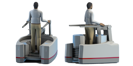
A new orthopedic CT system promises to improve the standard of care for knee imaging. On the CurveBeam LineUp, patients are scanned while standing upright and fully weight bearing.

Traditional CT and MR images are acquired in a non-weight bearing position, leading to “missed diagnoses of meniscal damage,” according to Dr. Neil Segal, who has been overseeing research efforts using a LineUp prototype, first at the University of Iowa and currently at the University of Kansas.

Although plain radiographs can be acquired while the patient is in a full weight-bearing position, the optimal degree of knee flexion and X-Ray beam tilt to best visualize the joint surface is person specific.

“Difficulty in reproducing the same view of the joint over time impairs ability to detect joint disease, and the 2D nature of radiographs makes these images of overlapping bony anatomy very insensitive for detecting abnormalities until there is advanced joint damage,” Dr. Segal said.

The LineUp was developed by CurveBeam, a Pennsylvania-based company that specializes in extremity cone beam CT systems for orthopedics. CurveBeam anticipates it will submit an application for and receive FDA 510(k) clearance for the LineUp in 2017. The LineUp will be on display at RSNA at Booth #8008 in the North Hall.

CurveBeam introduced the pedCAT, a bilateral weight bearing CT system dedicated to the foot and ankle, in 2012. Since then, the device has been added to the imaging services of hospital foot & ankle sections, orthopedic clinics and podiatry offices.

Like the pedCAT, the LineUp will provide isotropic, three-dimensional volumes of the anatomy with a high resolution output of between 0.2 mm and 0.3 mm slices. The LineUp will be the only cone beam CT system that can provide bilateral, weight bearing scans.

A study led by Dr. Segal focused on osteophytes, one structure linked to pain in people with knee osteoarthritis. Knees of community-dwelling adults with knee OA were imaged with MRI (reference), fixed-flexion radiographs, and weight bearing CT. The sensitivity and accuracy for detecting osteophytes and subchondral cysts were higher with weight bearing CT imaging in comparison to fixed-flexion radiographs. The study was published in the August 2016 issue of the Journal of Orthopedic Research.

“Clinically, this is a highly meaningful improvement,” Dr. Segal said. “It suggests that weight-bearing CT could replace radiographs as the recommended means of assessing knee OA. This advancement is even more significant given that it was made without significantly increasing the radiation dose (0.01 mSv for SCT vs. 0.005–0.102 mSv for a series of knee radiographs).”

Another research effort led by Dr. Segal indicates weight-bearing CT arthrography studies can provide outstanding delineation of articular cartilage with better differentiation between cartilage and subchondral bone then MRI studies, while also visualizing the cruciate ligaments. In knees with osteoarthritis, meniscal tears not visualized on MRI were detectable on weight-bearing CT.

“Thus, we found that some potential advantages of weight-bearing CT over non-weight-bearing MRI/MRA include 3D measures of meniscal position, detection of pathology not detected in unloaded positions, and ability to bear weight in a functional position, thus better recreating the magnitude of body weight and muscle forces acting around the knee during usual standing,” Dr. Segal said.

Los Angeles Orthopedic Surgeon Scheduled to Perform First Bone Anchored Prosthesis Surgery in Southern California

Los Angeles, CA (PRWEB) September 19, 2016

Dr. Daniel C. Allison, a board-certified orthopedic surgeon in Los Angeles specializing in orthopedic oncology, anterior hip replacement, and joint reconstruction, has announced that he is scheduled to perform the first bone anchored (osteointegrated) prosthesis surgery in Southern California. The patient is a former police officer with an above-the-knee amputation.

“We are very excited to be the first practice in Southern California to offer the surgery,” said Dr. Allison. “We make limb preservation a priority for our soft tissue and bone sarcoma patients, and are constantly working to bring the latest surgical innovation techniques to our joint replacement and hip arthroplasty treatments for both adult and pediatric patients.”

Traditional prosthetics use a ball and socket design. The socket is placed around the end of the affected limb, to which the prosthetic arm or leg is then attached. While this approach is highly effective in many cases, it requires enough surface area on the remaining limb to adequately attach the cap with the socket. It can also present issues with comfort and fit.

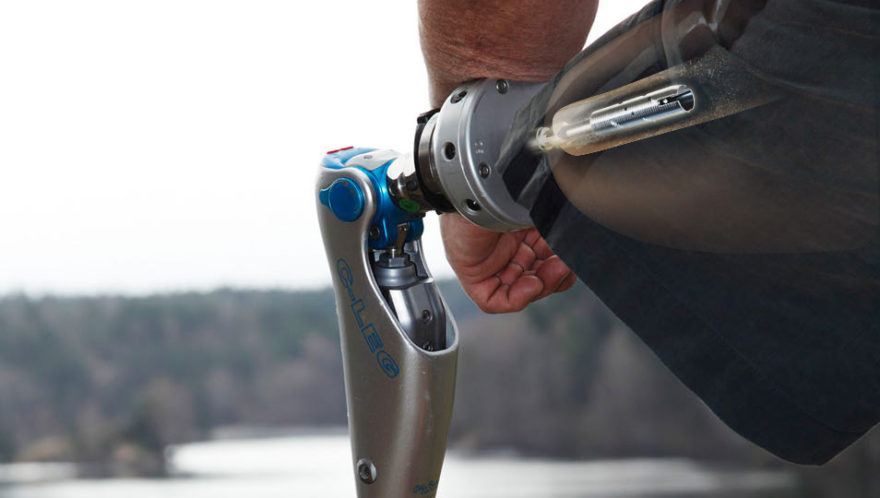
The Osseoanchored Prosthesis for the Rehabilitation of Amputees (OPRA), which has been approved by the FDA (Food and Drug Administration) for use in above the knee amputees, implants a titanium fixture directly into the bone. Once the fixture has integrated and healed, a rod is attached through the skin, which then connects to the prosthetic leg. The benefits of this approach include greater mobility, range of motion, and comfort. It also offers hope for amputees who are not good candidates for or have had limited success with traditional socket prosthetics. Using a titanium implant helps to mitigate the risk of rejection from the bone.

Learn about bone anchored prosthesis: http://www.DrAllison.org/Bone-Anchored-ProstethisOsteointegrated-OI-Prosthesis/

“With proper care and comprehensive physical therapy and rehabilitation, we are very optimistic that this approach will offer a significant improvement in quality of life for many adults, especially veterans and members of law enforcement like our patient, with above-the-knee amputations,” added Dr. Allison.

About Daniel C. Allison, MD, FACS.

Daniel C. Allison, MD, FACS, MBA, is board certified in orthopedic surgery with expertise in musculoskeletal oncology, joint reconstruction, and anterior hip replacement. He is Assistant Director of Orthopedic Oncology, Cedars-Sinai Sam Oschin Cancer Center. Dr. Allison is a fellow of the American College of Surgeons and the American Academy of Orthopedic Surgeons. As an orthopedic oncologist, Dr. Allison is a recognized leader in malignant problems of the musculoskeletal system, including soft tissue and primary sarcomas of the bone, invasive metastatic bone cancer, and invasive skin cancers of the back, pelvis, shoulder girdle, and extremities.

For more information about us, please visit http://www.DrAllison.org/

Daniel C. Allison, MD FACS
444 S San Vicente Blvd #603
Los Angeles, CA 90048
info(at)DrAllison(dot)org

Integra LifeSciences Announces First Clinical Uses of Cadence™ Total Ankle System in Europe

Plainsboro, N.J., Sept. 20, 2016 (Globe Newswire)

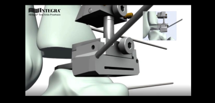
(NASDAQ:IART), a leading global medical technology company, today announced that the first three procedures using the Integra® Cadence™ Total Ankle System in Europe were successfully completed since July 13, 2016 by Dr. Thibaut Leemrijse, of the Foot and Ankle Institute, Parc Léopold Clinic, Brussels, Belgium. Internationally renowned for his expertise in foot surgery, Dr. Leemrijse and his team are considered leaders in the field of ankle arthroplasty.

“Integra’s new Cadence system offers a two-component implant, designed to adapt to a patient’s anatomy by providing improved range of motion, intraoperatively,” said Dr. Leemrijse. “The instrumentation allows ideal positioning of the implant, and the learning curve for this new device seems short, based on our first cases.”

The European introduction of the Cadence system supports Integra’s role as a leader in this rapidly expanding market. New and improved designs and advanced surgical education programs, such as the ones developed by Integra, are fueling the growth of the ankle prosthesis segment.

“Our innovative arthroplasty device is an example of the close cooperation between our company and expert surgeons, which seeks to develop devices that can be efficiently implanted in various anatomies,” said Stéphane Corp, Integra’s Vice President, Orthopedics & Tissue Technologies, for Europe, Middle East and Africa.

The Cadence system’s differentiated anatomic design and streamlined surgical technique allow Integra to continue offering a complete set of options to treat the continuum of care in total ankle prosthesis, one of the fastest growing extremities segments.

About Integra
Integra LifeSciences, a world leader in medical technology, is dedicated to limiting uncertainty for clinicians, so they can concentrate on providing the best patient care. Integra offers innovative solutions, including leading plastic and regenerative technologies, in specialty surgical solutions, orthopedics and tissue technologies. For more information, please visit www.integralife.com.

This news release contains forward-looking statements within the meaning of the Private Securities Litigation Reform Act of 1995. Forward-looking statements include, but are not limited to, statements concerning the products and services provided by Integra. Such forward-looking statements involve risks and uncertainties that could cause actual results to differ materially from predicted or expected results. Among other things, the willingness of surgical professionals to use Integra products may affect the prospects for their use in surgical procedures. In addition, the economic, competitive, governmental, technological and other factors, identified under the heading “Risk Factors” included in Item IA of Integra’s Annual Report on Form 10-K for the year ended December 31, 2015 and information contained in subsequent filings with the Securities and Exchange Commission could affect actual results.


CONTACT: Integra LifeSciences Holdings Company

Investors
Angela Steinway
609-936-2268
angela.steinway@integralife.com

Michael Beaulieu
609-750-2827
michael.beaulieu@integralife.com

Media Europe
Marie-Anne Girel
marie-anne.girel@integralife.com

Media U.S.
Gianna Sabella
609-775-8553
gianna.sabella@integralife.com

3D Printing Helps Surgeon Restore Child’s Sporting Ambitions and Reduce Surgery Time from 4 hours to Under 30 minutes

BELFAST (PRWEB UK) 13 SEPTEMBER 2016

When anxious parents approached Mr Michael Eames, Consultant Orthopaedic Surgeon at the South Eastern Trust, to investigate and help correct their young son’s injured forearm, little did they know that 3D printing would so drastically change the course of their son’s treatment.

Having suffered a complex forearm injury as a toddler a number of years earlier, the young patient’s bones had healed in an abnormal position preventing him from rotating his arm. This restriction of movement in a young child can be very detrimental to his development, sporting ambitions and career aspirations.

According to Michael Eames, the patient’s CT scan confirmed that the bones had, as suspected, healed incorrectly leading the team down the standard line of treatment of trying to reshape the bones to gain normal rotation. However, having access to a full-scale 3D printed model of the patient’s bones changed everything.

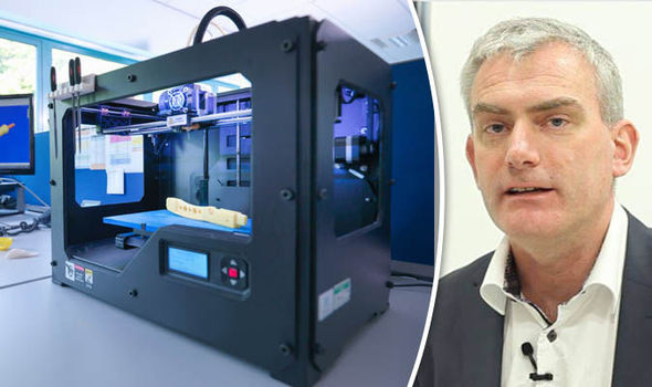
The South Eastern Trust supplied the patient’s CT scan data to Belfast-based medical 3D printing firm, Axial3D that produces bespoke hard and soft tissue models from 2D patient scan data for public and private healthcare across a wide variety of specialisms including orthopaedics, oncology, maxillofacial, cardiac and trauma.

Michael Eames said that the 3D printed model of his patient’s forearm bones offered unprecedented insight for diagnosing his patient and ultimately changed the course of his treatment, “We approached Axial3D to have the bones printed and that was enlightening. It showed us that while the shape of the bones was grossly abnormal, it wasn’t the shape of the bones that was limiting the rotation. It was the tight structures between the bones that had scarred down that was preventing the child from rotating his arm. Having a tangible scale piece of anatomy provides huge insight into the pathology not possible on conventional CT or MRI scans.”

Access to a bespoke 3D printed model of the patient’s forearm bones changed the standard course of treatment from a 4 hour complex surgery to reshape the bones and the ensuing rehabilitation, to a much less evasive soft tissue procedure that took just under 30 minutes.

According to Daniel Crawford, Axial3D CEO, their bespoke models offer clinicians a deeper appreciation of their patient’s ailment or injury beyond what is possible with conventional 2D imaging: “Axial3D’s patient-specific anatomical models give clinicians greater insight and therefore confidence in diagnosing and treating complex cases. In some cases, our models have been used to develop and trial novel surgical techniques pushing the boundaries of surgical intervention. The adoption of 3D medical printing has the potential to revolutionise medical provision.”

To fulfil its mission to provide easy access to patient-specific tools, Axial3D recently launched an online service for clinicians to upload patient CT and MRI scans, visualise them in 3D and order precise 3D printed models in minutes. For this young patient, access to the model provided a more accurate diagnosis, less evasive procedure, faster recovery and, four weeks post op, he was discharged with 90% range of movement in his forearm and no pain.

This week Axial3D is showcasing its bespoke 3D printed orthopaedic models at the British Orthopaedic Association Congress [13-16 September at the Waterfront Hall, Belfast], an annual event that is expected to attract over 1,000 orthopaedic surgeons to the city.

Here’s How to Convince the Brain That Prosthetic Legs Are Real

By  – September 12, 2016

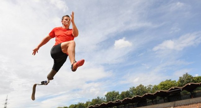
The carbon fiber legs or “blades” used by lower limb amputee runners have arguably become one of the most iconic symbols of the Paralympic Games. Although different lower-limb sports prostheses are used for running, jumping, and other activities, they share a single common aim: they are designed to help paralympians run faster and jump higher or further than other competitors. Form follows function.

For those who have prostheses for more everyday uses, however, their replacement limbs need to be able to adapt to different scenarios and perform a variety of functions, not just excel in one discipline — just like an actual leg. So how can we make prostheses feel more like the real thing rather than a specialist tool?

Whereas modern running blades have a distinctive hook shape, one of the most promising engineering approaches for everyday prostheses is to closely model the biological design of a leg, ankle and foot. This approach is referred to as “biomimicry.”

A “passive” ankle-foot prosthesis generally uses elastic like a spring to replicate the behavior of the Achilles tendon, storing elastic energy and releasing it before ankle push-off. “Active” prostheses additionally use an actuator or motor to make up for the power previously provided by the calf muscle at every step. Such prostheses have been shown to help users walk more like a non-amputee and improve symmetry between the biological and the artificial limb. At the moment, this mainly applies to walking overground at steady speeds rather than activities such as climbing stairs.

Simple design, advanced technology

Other ways to make a prosthesis more like a biological leg and improve the user’s comfort are more simple. They also illustrate how important it is to involve the amputee in the design process. One user of the most advanced bionic ankle currently available told me its greatest feature was not that it provided a powered push-off or that it allowed them to walk more like a non-amputee. Instead, it was that the foot dropped flaton the ground when sitting with an outstretched leg, rather than sticking up awkwardly at a 90-degree angle (as is the case for the majority of prosthetic feet).


READ THE REST HERE